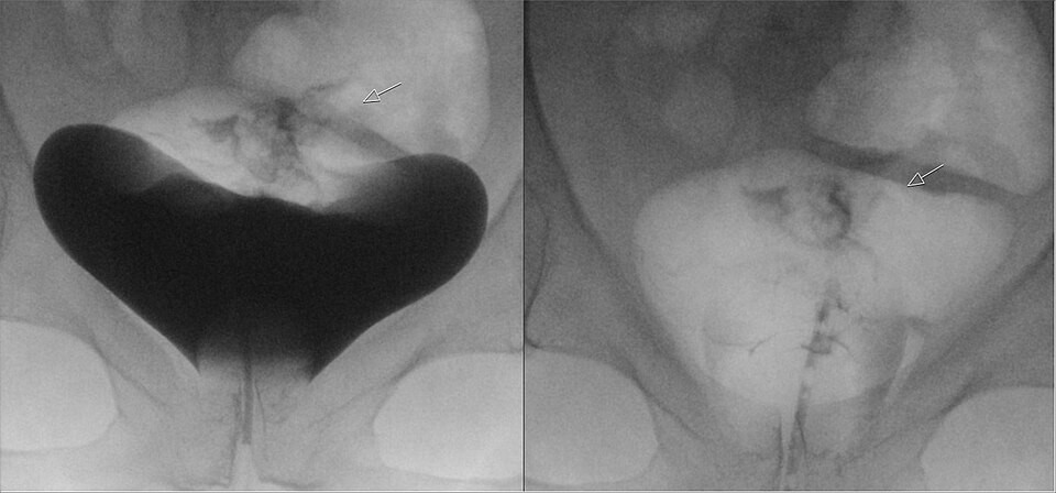
1. These AP images are from a retrograde cystogram on a patient with a pelvic fracture. The left image was taken with the bladder distended, and the right image after the bladder was drained. Why is the drained image part of the examination?
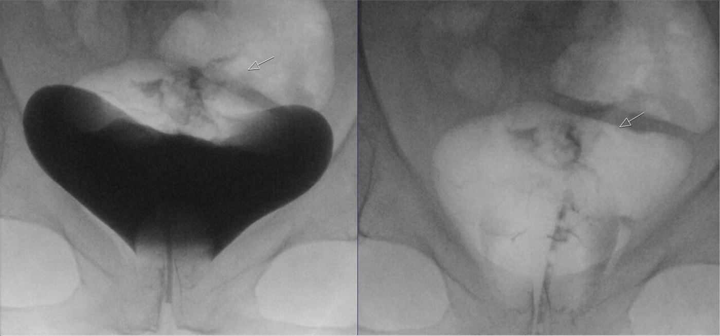

A retrograde cystogram fills the bladder with contrast through a urethral catheter. A voiding cystourethrogram (VCUG) adds images while the patient urinates, to show the urethra and any reflux of urine up the ureters. Many VCUGs are done on children.
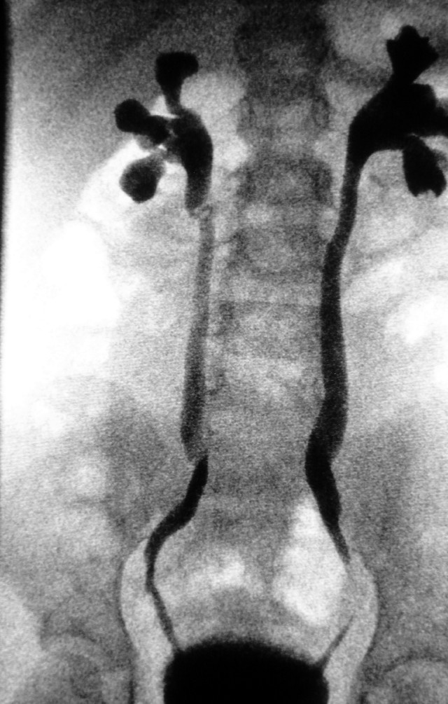
1. These AP images are from a retrograde cystogram on a patient with a pelvic fracture. The left image was taken with the bladder distended, and the right image after the bladder was drained. Why is the drained image part of the examination?
2. For the AP projection during retrograde cystography, how is the CR angled to project the pubic bones below the bladder?
3. During a voiding cystourethrogram on a 2-year-old, the radiologist asks the radiographer to remove the grid from the fluoroscopic receptor. What is the rationale?
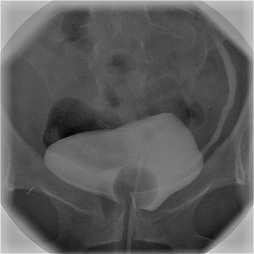
4. This cystogram shows a contrast-filled urinary bladder with a catheter entering from below. How does contrast reach the bladder in a retrograde cystogram?
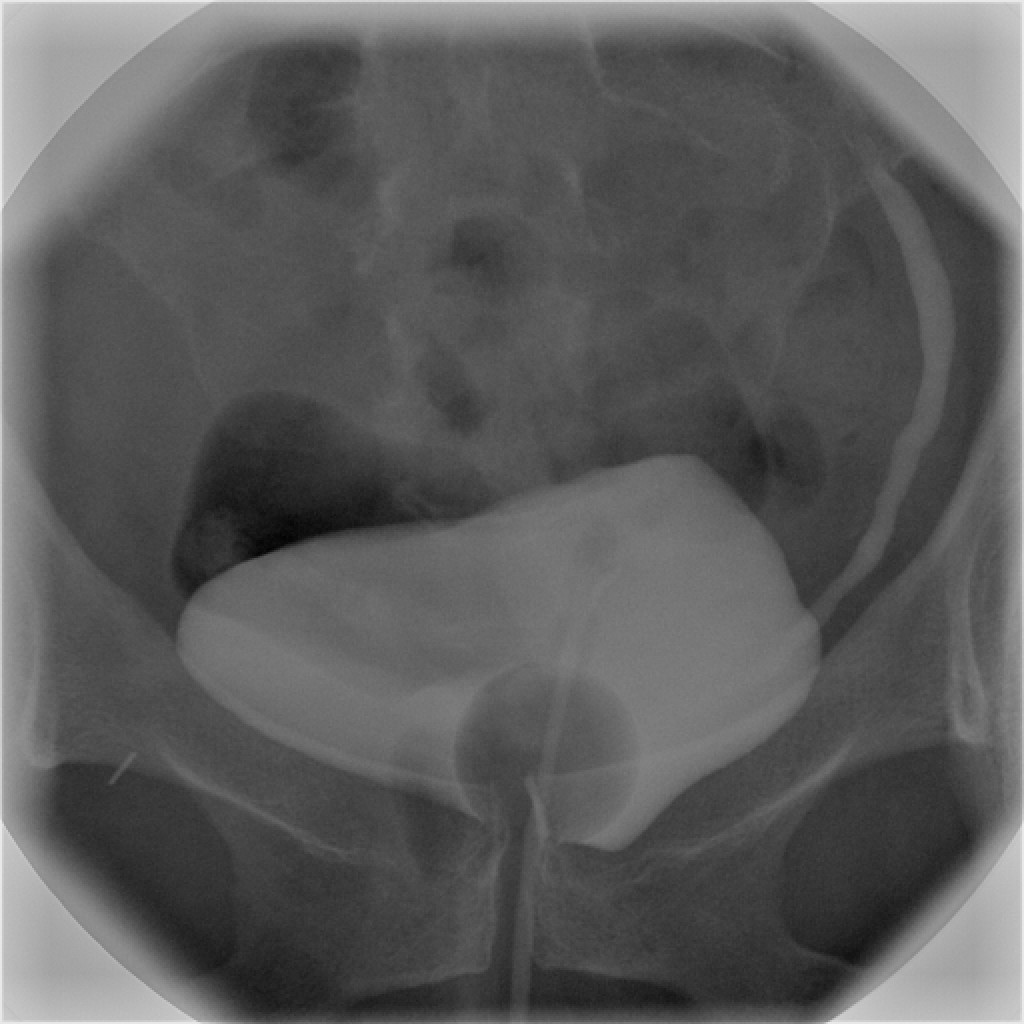
5. This cystogram after bladder repair shows the filled bladder (left) and the image after drainage (right), where contrast remains outside the bladder outline at the top. What does this show?
The parts, the movements, the dose modes, and where to stand.
What's sterile, what you can touch, and how to move around the field.
Barium swallow and UGI: why they're ordered, the positions, and your role.
Video swallow study with speech therapy: bolus types, the field of view, and your role.
Timed films as barium moves from the stomach to the colon.
Single- and double-contrast BE: prep, tip insertion, positions, and overheads.
Uterus and fallopian tubes for infertility: timing, setup, and dose.
Joint injection under fluoro, usually before MRI or CT.
Contrast in the spinal canal: needle site, table tilt, and keeping contrast out of the head.
Spinal tap under fluoro when bedside attempts fail.
Bile ducts through a scope, in surgery, or through a T-tube.
The most common C-arm case: the fracture table, the two views, and your role.
Open reduction and internal fixation of the ankle or wrist: the views, the mini C-arm, and hands in the beam.