
1. Shortly after IV iodinated contrast is injected for a fluoroscopic venogram, the patient develops a few scattered hives. Breathing is easy, the voice is normal, and blood pressure is 128/76 mm Hg. Which response is MOST appropriate?
Arthrogram
An arthrogram is an injection of contrast into a joint under fluoroscopy. Today most arthrograms are the first step of an MR or CT arthrogram: the radiologist places the needle and injects under fluoro, then the patient goes straight to MRI or CT. The shoulder, hip, wrist and knee are the most common joints.
What the ARRT tests
- Sterile procedure with written informed consent; skin prep and a local anesthetic.
- Contrast: iodinated contrast for fluoro and CT; a diluted gadolinium mix for MR arthrography; air may be added for a double-contrast study.
- Contraindications: infection at the injection site or in the joint, and allergy to the contrast used.
- Move the joint after the injection to spread the contrast, then image soon, because the contrast is absorbed and loses sharpness over time.
- Knee arthrogram (rare now): stress views of the medial and lateral menisci. Wrist: the triangular fibrocartilage complex (TFCC). Shoulder: rotator cuff and labrum.
Why it's done
- Suspected tear of the rotator cuff, labrum (shoulder or hip), cartilage, ligament or meniscus.
- Checking a joint after surgery or a joint replacement for loosening.
- Joint aspiration: sampling fluid for infection.
Who's in the room
- Radiologist
- Gets consent, preps the skin, places the needle under fluoro, and injects.
- Radiographer (you)
- Sets up the sterile tray and contrast, positions the joint, runs the fluoro, and moves the patient to MRI or CT afterward.
Step by step
- Before. Confirm the order, allergies, blood thinners per protocol, and pregnancy status. Prepare the sterile tray, local anesthetic, needles, extension tubing and contrast mix.
- Position. Position the joint for the needle path, for example shoulder supine with the arm externally rotated, hip supine with the leg slightly internally rotated.
- Injection. The radiologist preps the skin, numbs it, places the needle under fluoro, confirms the tip in the joint with a little contrast, then injects the full dose.
- After. Bandage, have the patient move the joint gently, and take the patient to MRI or CT without a long wait. Explain that the joint may feel full or sore for a day.
Your job
- Sterile field: don't reach over it; open supplies onto it correctly.
- Know which contrast mix your radiologist uses for MR vs CT arthrograms.
- Coordinate with MRI or CT so the scan starts soon after the injection.
Watch out for
- Infection: report fever, redness or worsening pain after the exam.
- MR safety: screen the patient for MRI before the injection, not after.
- Vasovagal reactions are common with needles; keep the patient lying down.
Images
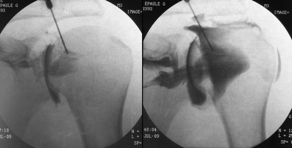
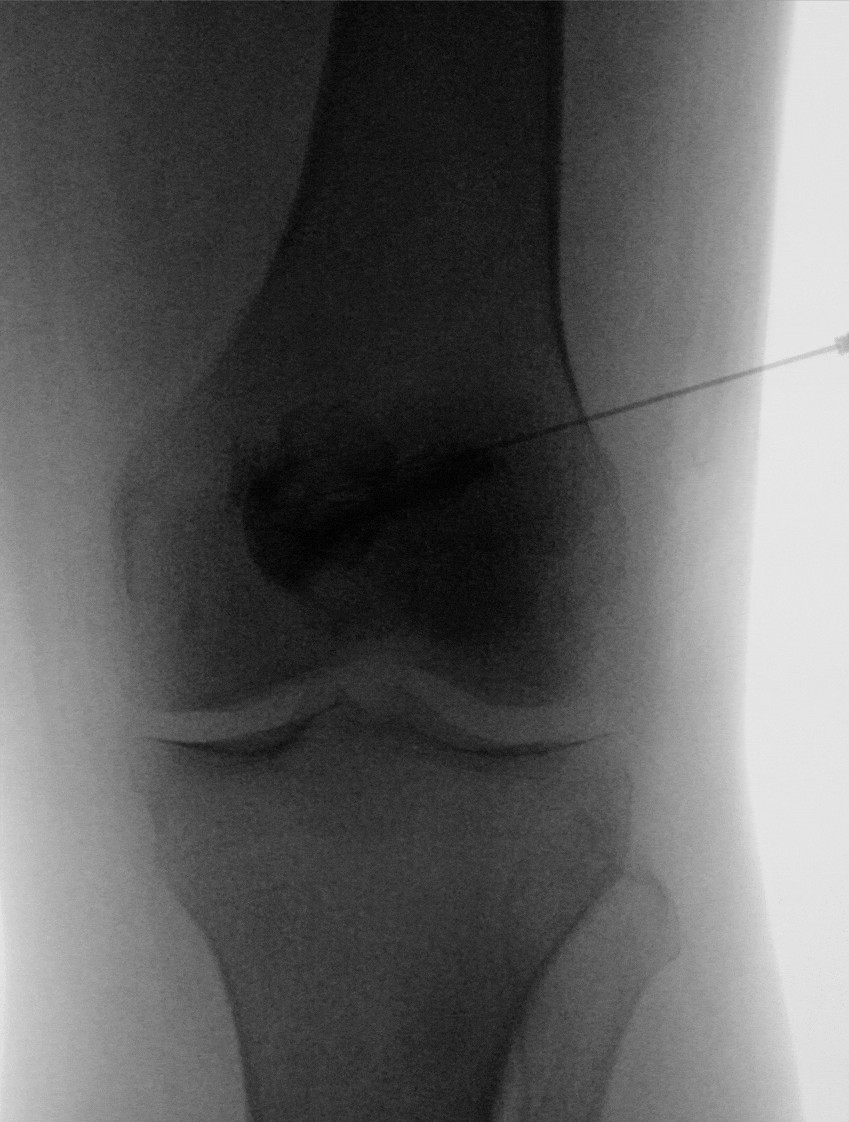
Quick check
2. During a fluoroscopic arthrogram, which action by a nonsterile team member breaks sterile technique?
3. Why are images obtained promptly after contrast is injected for a conventional arthrogram?
4. Before an arthrogram or another invasive procedure, the team pauses to confirm the patient, procedure and site. What is this step called?
5. A 15-year-old who is not an emancipated minor is scheduled for a hip arthrogram and arrives with an adult family friend who is not a legal guardian. What should the radiographer do?
Practice it in 3D
More in the guide
C-arm basics
The parts, the movements, the dose modes, and where to stand.
Sterile basics
What's sterile, what you can touch, and how to move around the field.
Esophagram and upper GI series
Barium swallow and UGI: why they're ordered, the positions, and your role.
Modified barium swallow (swallow study)
Video swallow study with speech therapy: bolus types, the field of view, and your role.
Small bowel follow-through
Timed films as barium moves from the stomach to the colon.
Barium enema
Single- and double-contrast BE: prep, tip insertion, positions, and overheads.
Cystogram and VCUG
Bladder filled through a catheter: leak checks, reflux, and voiding images.
Hysterosalpingogram (HSG)
Uterus and fallopian tubes for infertility: timing, setup, and dose.
Myelogram
Contrast in the spinal canal: needle site, table tilt, and keeping contrast out of the head.
Lumbar puncture (fluoro-guided)
Spinal tap under fluoro when bedside attempts fail.
ERCP and cholangiograms
Bile ducts through a scope, in surgery, or through a T-tube.
Hip pinning (hip fracture fixation)
The most common C-arm case: the fracture table, the two views, and your role.
Ankle and wrist ORIF
Open reduction and internal fixation of the ankle or wrist: the views, the mini C-arm, and hands in the beam.