RAD Registry Prep logo

Fluoro & OR Guide

Hip pinning (hip fracture fixation)

Hip fracture fixation is one of the most common C-arm cases. The patient is usually elderly and broke the hip in a fall. The surgeon fixes it with screws, a sliding hip screw with a side plate, or a nail down the femur, and needs a clean AP and lateral of the hip at every step.

What the ARRT tests

  • A shortened, externally rotated leg suggests a hip fracture. Don't rotate the leg for the AP.
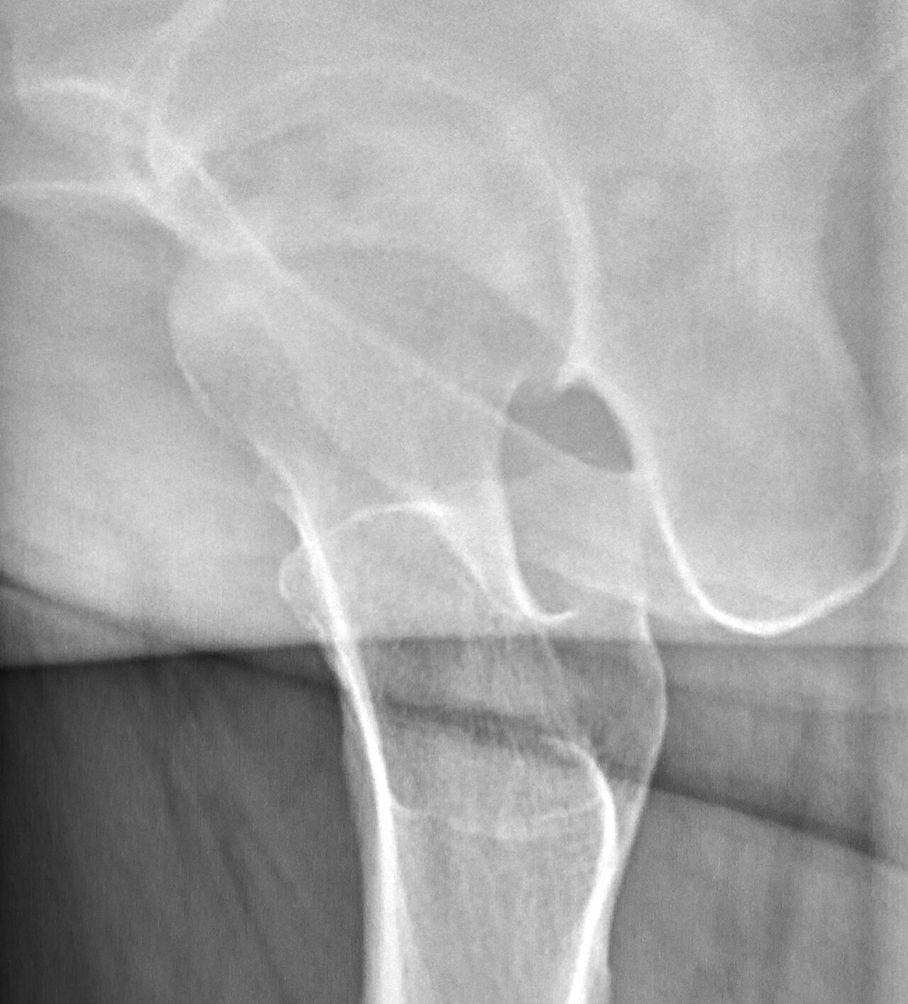
  • The frog-leg lateral is never used when a fracture is suspected; the trauma lateral is the cross-table axiolateral (Danelius-Miller).
  • Mobile fluoroscopy: tube under the patient, receptor close, collimate, pulsed fluoro, and stand on the receptor side.
  • Surgical asepsis around the sterile field.

Why it's done

  • Femoral neck or intertrochanteric fracture, usually from a fall in an older patient, often with osteoporosis.
  • Symptoms: hip or groin pain, can't bear weight, leg shortened and externally rotated.
  • Nondisplaced neck fractures: cannulated screws. Intertrochanteric: sliding hip screw or a cephalomedullary (intramedullary) nail. Displaced neck fractures in older patients often get a partial or total hip replacement instead, with less fluoro.

Who's in the room

Orthopedic surgeon and assistant
Reduce the fracture, place the guide wire, ream and place the hardware, and call for images.
Anesthesia
Keeps the patient asleep or sedated at the head of the bed.
Scrub tech
Runs the instruments and implants, and drapes the C-arm with you.
Circulating nurse
Non-sterile; handles the room, supplies and paperwork, and helps you move the C-arm in.
Radiographer (you)
Runs the C-arm, gets the AP and lateral on command, saves images, and keeps radiation exposure down.

Step by step

  1. Positioning. The patient goes on a fracture (traction) table: the broken leg in a traction boot, the good leg raised out of the way in a leg holder. The C-arm usually comes in from the good side, between the legs.
  2. Before draping. Get the surgeon's AP and lateral of the hip. The surgeon pulls traction and rotates the leg to reduce the fracture while you show it live. Note your angles or tape the floor.
  3. Draping. The patient and the C-arm are draped. From here on, every move of the C-arm must keep the drape clear of non-sterile surfaces.
  4. Guide wire. The surgeon drills a guide wire into the femoral head. You alternate AP and lateral so they can see the wire is centered in the head on both views.
  5. Hardware. Reaming, the screw or nail, and the side plate or locking screws, each checked with images.
  6. Final images. Final AP and lateral saved and sent to PACS. Record fluoro time and dose.

Your job

  • Know your two positions: an AP (tube under the table) and a lateral (orbital rotation about 90°, angled to follow the femoral neck).
  • Move between AP and lateral fast and smoothly, without hitting the table or the good leg.
  • Label images right or left as your department requires, and save the ones the surgeon asks for.
  • Keep collimation tight and use pulsed mode; the lateral hip is a high-dose view.

Watch out for

  • Before draping, make sure you can get a full lateral. If the good leg or the table is in the way, fix it now, not after the patient is prepped.
  • The drape snagging on the table or the good leg during the swing to lateral.
  • Large patients: the lateral needs a lot of technique. Bring the receptor in close, stand back during shots, and tell the surgeon if the image can't be improved.
  • Fragile, elderly skin and pressure points on the fracture table.

Tips from the room

  • Ask the surgeon before draping how they like the images shown (flipped or rotated) and set it once.
  • Many surgeons say "shoot" for a single shot and "live" for continuous fluoro. Learn their words on the first case.
  • Keep the C-arm parked out of the way between steps, but close enough to get back to position fast.

Images

Hip fracture before fixation.
Cross-table (axiolateral) hip: the lateral view used in trauma.

Quick check

1. This AP hip radiograph of a patient in the emergency department shows a displaced, comminuted proximal femur fracture. The patient can flex and raise the unaffected leg. Which lateral projection should the radiographer perform?
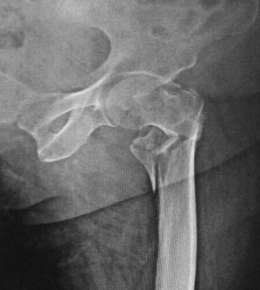

Image: Memon, Patel & Juva. CC BY 4.0.

2. A mobile C-arm has a source-to-image distance (SID) of 100 cm. The patient's abdomen measures 32 cm, and the image intensifier is 40 cm above the patient's anterior skin. Ignoring the table, what is the source-to-skin distance, and is it acceptable?

3. During a C-arm hip pinning, the radiographer rotates the C-arm from the AP position to a lateral projection through the hip. With automatic brightness control active, what happens to the patient's dose rate?

4. During a C-arm hip pinning, a large metal plate and screws fill the center of the field. The displayed kVp and mA rise sharply, and the bone and soft tissue around the hardware appear washed out. What best explains this?

5. For the axiolateral inferosuperior (cross-table) hip, how is the unaffected leg positioned?

Image: Mikael Häggström, M.D. CC0 1.0.

Practice it in 3D

More in the guide