
1. A patient with a suspected herniated nucleus pulposus is scheduled for lumbar myelography. Into which space is the contrast medium injected?
Myelogram
A myelogram puts contrast into the subarachnoid space around the spinal cord and nerve roots through a spinal needle, then images the spine under fluoro and usually with CT right after. MRI has replaced most myelograms, but they're still done when a patient can't have an MRI.
What the ARRT tests
- Contrast: nonionic, water-soluble iodinated contrast approved for intrathecal use. Never use a product that isn't labeled for intrathecal injection.
- Needle site: usually L2–L3 or L3–L4 (below the end of the spinal cord at about L1–L2). A lateral C1–C2 puncture is an alternate site.
- Patient prone with a pillow under the abdomen to flex the lumbar spine and open the spaces.
- The table tilts to move the contrast: head down to send it to the thoracic or cervical spine. The head is kept hyperextended (chin up) so the contrast doesn't flow into the head.
- Horizontal beam (cross-table) laterals are taken with the patient prone.
- Contraindications: blood in the CSF, increased intracranial pressure, and a recent lumbar puncture (within about 2 weeks). Some medicines that lower the seizure threshold, and blood thinners, are held per protocol.
- After: head elevated, fluids, and rest; headache is the most common side effect.
Why it's done
- Spinal stenosis, nerve root compression or a disk herniation in a patient who can't have an MRI (some pacemakers and implants, severe claustrophobia).
- Metal hardware that makes MRI images hard to read.
- Suspected CSF leak.
Who's in the room
- Radiologist
- Gets consent, places the needle, injects the contrast and tilts the patient to move it.
- Radiographer (you)
- Sets up the tray and contrast, positions the patient, runs the fluoro, tilts the table on command, and takes the cross-table laterals.
- CT technologist
- Scans the spine right after the fluoro portion (post-myelogram CT).
Step by step
- Before. Confirm consent, allergies, medications (blood thinners, seizure-threshold drugs), kidney function if required, and pregnancy status. Footboard and shoulder supports on the table.
- Puncture. Patient prone with a pillow under the abdomen. The radiologist places the needle under fluoro. CSF may be collected; label the tubes in order.
- Contrast. Contrast is injected slowly. The radiologist tilts the table to move it to the area of interest while images are taken, including cross-table laterals.
- After. Keep the head up when moving the patient. Transfer to CT. Explain the aftercare: head elevated, plenty of fluids, and rest for the rest of the day.
Your job
- Table safety: footboard and shoulder supports before any tilt.
- Keep the head extended during head-down tilt.
- Have the contrast label checked by the radiologist (the product must be approved for intrathecal use).
Watch out for
- Contrast moving toward the head: sit the patient up gently if told; keep the head elevated.
- Seizures, severe headache, or numbness: tell the radiologist right away.
- Falls when the patient stands up after lying prone; move slowly.
Images
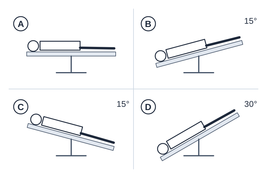
Quick check
2. A child is placed in the lateral recumbent position for a lumbar puncture, and the knees are drawn up toward the chest with the head bent forward. Why is the lumbar spine flexed for this procedure?

3. A radiographer is assisting with a sterile tray for a fluoroscopic lumbar puncture. Which practice maintains the sterile field?
4. Which task is within the radiographer's role during a fluoroscopically guided lumbar puncture for myelography?
5. While setting up for a lumbar myelogram, the radiographer finds that the only iodinated contrast on the cart is an ionic agent labeled for intravascular use. What should the radiographer do?
Practice it in 3D
More in the guide
C-arm basics
The parts, the movements, the dose modes, and where to stand.
Sterile basics
What's sterile, what you can touch, and how to move around the field.
Esophagram and upper GI series
Barium swallow and UGI: why they're ordered, the positions, and your role.
Modified barium swallow (swallow study)
Video swallow study with speech therapy: bolus types, the field of view, and your role.
Small bowel follow-through
Timed films as barium moves from the stomach to the colon.
Barium enema
Single- and double-contrast BE: prep, tip insertion, positions, and overheads.
Cystogram and VCUG
Bladder filled through a catheter: leak checks, reflux, and voiding images.
Hysterosalpingogram (HSG)
Uterus and fallopian tubes for infertility: timing, setup, and dose.
Arthrogram
Joint injection under fluoro, usually before MRI or CT.
Lumbar puncture (fluoro-guided)
Spinal tap under fluoro when bedside attempts fail.
ERCP and cholangiograms
Bile ducts through a scope, in surgery, or through a T-tube.
Hip pinning (hip fracture fixation)
The most common C-arm case: the fracture table, the two views, and your role.
Ankle and wrist ORIF
Open reduction and internal fixation of the ankle or wrist: the views, the mini C-arm, and hands in the beam.