
1. During hysterosalpingography, contrast fills the uterus. The right uterine tube spills freely into the peritoneal cavity, but the left tube fills to a rounded, dilated end with no spill. What do these findings indicate?
Hysterosalpingogram (HSG)
The hysterosalpingogram (HSG) shows the inside of the uterus and whether the fallopian tubes are open. Contrast is injected through the cervix while the radiologist or gynecologist watches under fluoro. You set up the sterile tray, position the patient and keep the dose low, since the ovaries are in the beam.
What the ARRT tests
- Timing: about 7–10 days after the first day of the menstrual period, after bleeding stops and before ovulation, so the patient is not pregnant.
- Contraindications: pregnancy, active pelvic infection, and active bleeding.
- Water-soluble iodinated contrast is injected through a cannula or balloon catheter in the cervix.
- Patent tubes: contrast spills freely from the ends of the tubes into the peritoneal cavity.
- Patient supine, often in the lithotomy position; the uterus and tubes are near the center of the pelvis.
- The ovaries are in the beam, so pulsed fluoro, last-image hold and tight collimation matter.
Why it's done
- Infertility workup: blocked tubes, scarring, or a uterine shape problem (polyps, fibroids, a septate uterus).
- Checking tubal blockage after a sterilization procedure.
- Repeated miscarriages.
Who's in the room
- Radiologist or gynecologist
- Places the speculum and catheter, injects the contrast, and takes the spot images.
- Radiographer (you)
- Sets up the sterile tray and contrast, positions the patient, runs or assists with the fluoro, and protects the patient's privacy.
Step by step
- Before. Confirm the date of the last menstrual period and pregnancy test per protocol, allergies, and any pelvic infection. Explain the exam and that cramping is common. Many patients take an over-the-counter pain reliever beforehand.
- Setup. Patient supine, knees up and apart (lithotomy). Sterile tray with speculum, cannula or balloon catheter, cleansing solution, and contrast drawn up without air bubbles.
- Injection. The doctor places the catheter and injects slowly while watching: first the uterine cavity, then the tubes, then spill into the pelvis.
- After. Remove the instruments, give the patient a pad and privacy, and explain that light spotting and cramps are normal. Watch for signs of infection over the next days.
Your job
- Make sure the timing and pregnancy screening are documented before anyone comes in.
- Keep the room warm and private; this exam is uncomfortable and personal.
- Low dose: pulsed fluoro, last-image hold, tight collimation.
Watch out for
- Vasovagal reaction from cervical manipulation: dizziness, sweating, a slow pulse.
- Contrast reaction: rare, but know the allergy history.
- Air bubbles in the syringe can look like polyps; prepare the contrast carefully.
Quick check
2. This image was obtained during a hysterosalpingogram. Contrast has passed from the uterine cavity into a fallopian tube. Which finding confirms that a fallopian tube is patent?
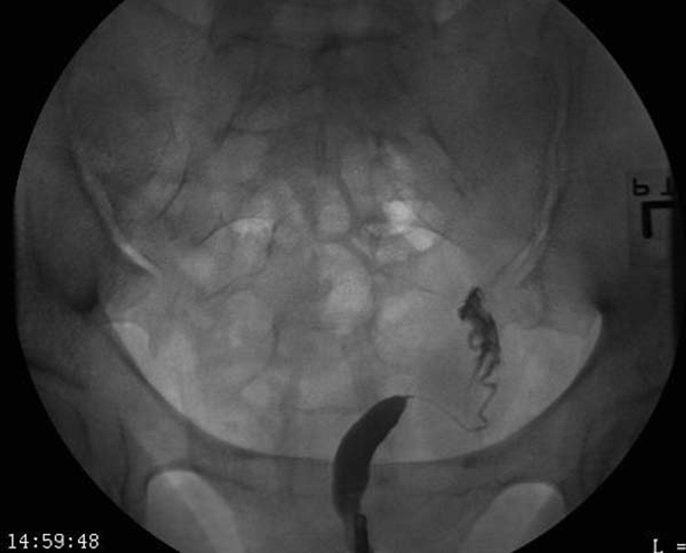
3. A patient scheduled for hysterosalpingography reports pelvic pain, fever and abnormal discharge, and the physician suspects an active pelvic infection. What is the appropriate action?
4. A patient is prepared for hysterosalpingography, and the radiologist will insert a speculum and cervical cannula. How should the patient be positioned on the fluoroscopic table?
5. What procedure is shown in this image?
Practice it in 3D
More in the guide
C-arm basics
The parts, the movements, the dose modes, and where to stand.
Sterile basics
What's sterile, what you can touch, and how to move around the field.
Esophagram and upper GI series
Barium swallow and UGI: why they're ordered, the positions, and your role.
Modified barium swallow (swallow study)
Video swallow study with speech therapy: bolus types, the field of view, and your role.
Small bowel follow-through
Timed films as barium moves from the stomach to the colon.
Barium enema
Single- and double-contrast BE: prep, tip insertion, positions, and overheads.
Cystogram and VCUG
Bladder filled through a catheter: leak checks, reflux, and voiding images.
Arthrogram
Joint injection under fluoro, usually before MRI or CT.
Myelogram
Contrast in the spinal canal: needle site, table tilt, and keeping contrast out of the head.
Lumbar puncture (fluoro-guided)
Spinal tap under fluoro when bedside attempts fail.
ERCP and cholangiograms
Bile ducts through a scope, in surgery, or through a T-tube.
Hip pinning (hip fracture fixation)
The most common C-arm case: the fracture table, the two views, and your role.
Ankle and wrist ORIF
Open reduction and internal fixation of the ankle or wrist: the views, the mini C-arm, and hands in the beam.