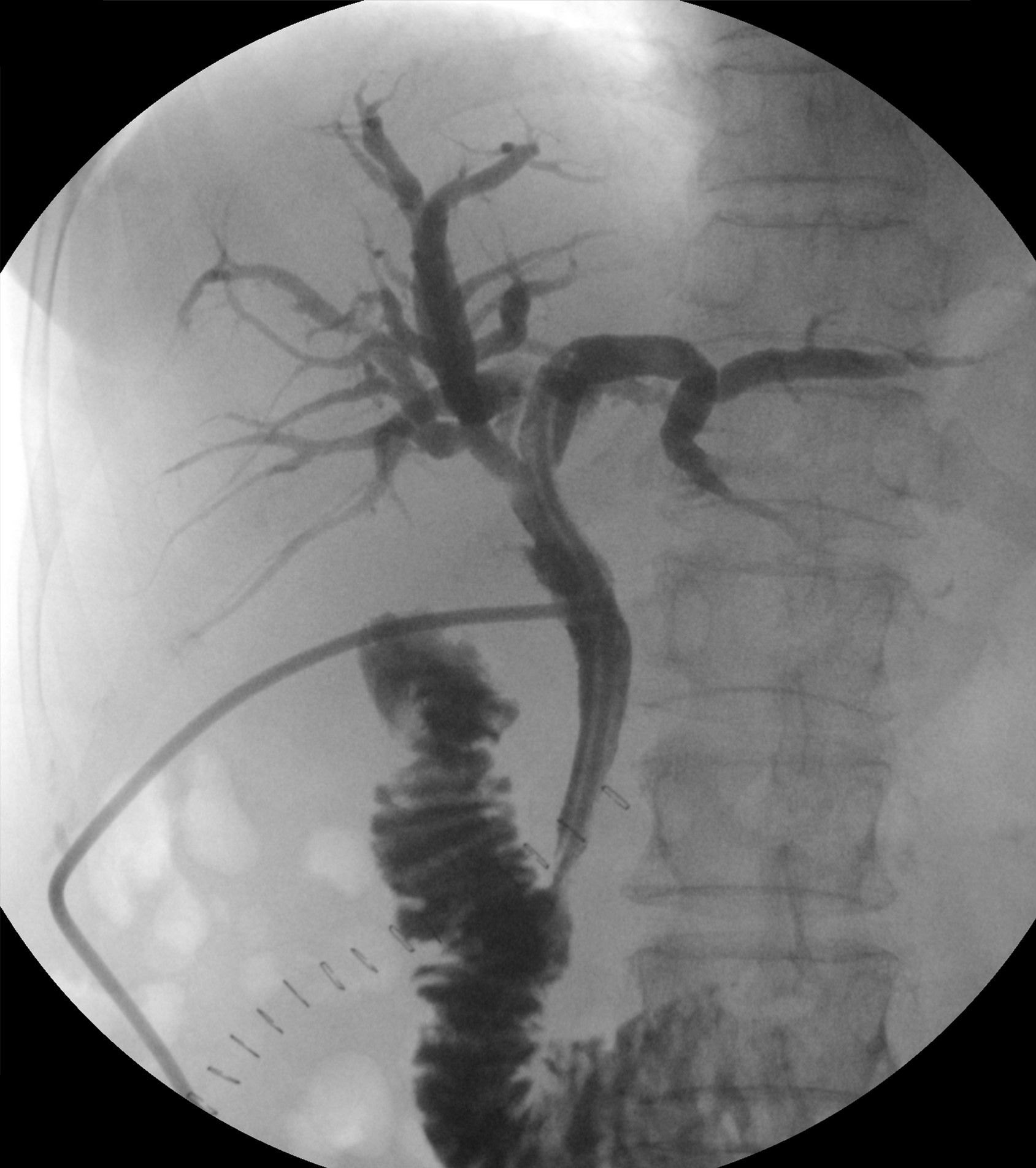
1. During an operative (surgical) cholangiogram, the C-arm is used in the AP position. Which C-arm orientation BEST reduces scatter to the head and neck of the surgical team?

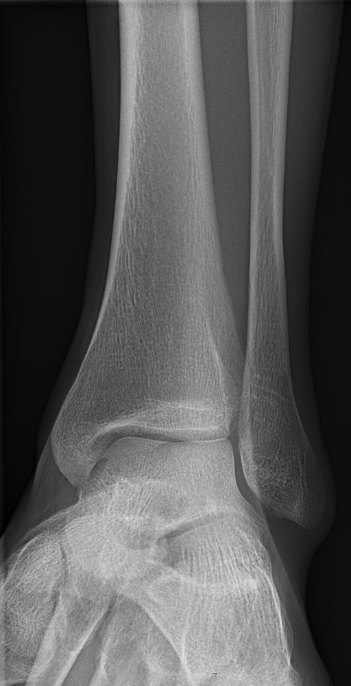
ORIF means open reduction and internal fixation: the surgeon opens the skin, puts the bones back in place, and holds them with plates and screws. Ankle and wrist fractures are the most common ORIF cases you'll run. They're often done with a mini C-arm.
1. During an operative (surgical) cholangiogram, the C-arm is used in the AP position. Which C-arm orientation BEST reduces scatter to the head and neck of the surgical team?
2. During a C-arm hip pinning, the radiographer wears a wraparound lead apron and stands about 2 m from the patient. The surgeon will operate the fluoroscopy footswitch for the next several minutes, and the radiographer is not needed at the C-arm. Which action BEST further reduces the radiographer's occupational dose?
3. Which ankle projection BEST demonstrates the entire mortise, with the medial, superior, and lateral joint spaces open and equal?
4. The operating room schedule lists four cases needing intraoperative fluoroscopy. For which case is a mini C-arm the appropriate unit?
5. During a lumbar interlaminar epidural steroid injection with the patient prone, the physician wants to judge how deep the needle tip has advanced. Which C-arm view is used?
The parts, the movements, the dose modes, and where to stand.
What's sterile, what you can touch, and how to move around the field.
Barium swallow and UGI: why they're ordered, the positions, and your role.
Video swallow study with speech therapy: bolus types, the field of view, and your role.
Timed films as barium moves from the stomach to the colon.
Single- and double-contrast BE: prep, tip insertion, positions, and overheads.
Bladder filled through a catheter: leak checks, reflux, and voiding images.
Uterus and fallopian tubes for infertility: timing, setup, and dose.
Joint injection under fluoro, usually before MRI or CT.
Contrast in the spinal canal: needle site, table tilt, and keeping contrast out of the head.
Spinal tap under fluoro when bedside attempts fail.
Bile ducts through a scope, in surgery, or through a T-tube.
The most common C-arm case: the fracture table, the two views, and your role.