RAD Registry Prep logo

Fluoro & OR Guide

Esophagram and upper GI series

The esophagram (barium swallow) looks at the esophagus. The upper GI series (UGI) looks at the distal esophagus, stomach and duodenum. The radiologist or radiologist assistant runs the fluoro; you set up, prepare the contrast, help the patient turn, and take the overhead images.

What the ARRT tests

  • Prep for a UGI: NPO about 8 hours (usually after midnight), no smoking or gum.
  • Barium is contraindicated if a perforation is suspected or before surgery; use a water-soluble iodinated contrast instead.
  • Double contrast: thick barium plus gas crystals coats the lining to show small lesions and ulcers.
  • Supine (and LPO): barium pools in the fundus, air rises to the body and antrum. Prone (and RAO): barium fills the body and pylorus, air rises to the fundus.
  • RAO esophagram: the esophagus shows between the spine and the heart.
  • Body habitus moves the stomach: high and transverse in a hypersthenic patient, low and vertical in an asthenic one.

Why it's done

  • Esophagram: trouble swallowing (dysphagia), pain with swallowing, food sticking, suspected stricture, ring or tumor, reflux, or a check after esophageal surgery.
  • UGI: heartburn and reflux (GERD), upper abdominal pain, suspected ulcer, hiatal hernia, nausea and vomiting, weight loss, or a leak check after bariatric surgery (often with water-soluble contrast).
  • A modified barium swallow (swallow study) is different: it's done with speech therapy to watch the swallow itself for aspiration.

Who's in the room

Radiologist or radiologist assistant
Takes the history, runs the fluoro, watches the swallow and the stomach in real time, and takes spot images.
Radiographer (you)
Sets up the room and contrast, prepares the patient, works the table and patient positions, takes overhead images, and manages radiation protection.
Patient
Swallows on command, holds their breath, and turns as asked, often while the table tilts.

Step by step

  1. Before. Confirm the order, NPO status, allergies, pregnancy status and history. Explain the exam. Set up the barium (thin and thick), gas crystals, cups and straws. Put the footboard on the table.
  2. Upright. With the table upright, the patient takes gas crystals and thick barium while the radiologist takes double-contrast images of the esophagus and stomach.
  3. Table down. The table goes horizontal. The patient turns (often a full roll) to coat the stomach, then holds positions for the radiologist's spots: the supine/LPO views for the air-contrast body and antrum, prone/RAO for barium-filled views and the duodenal bulb.
  4. Overheads. After fluoro, you take the overhead images your protocol lists (for example PA, RAO, right lateral and LPO), centered for the patient's body habitus.
  5. After. Tell the patient to drink plenty of fluids; barium can cause constipation and will make the stool white for a day or two. Record fluoro time.

Your job

  • Room, contrast and table ready before the patient walks in; footboard on.
  • History, allergies, NPO, pregnancy screening, and a clear explanation for the patient.
  • Help the patient turn safely on a narrow tilting table; keep a hand ready.
  • Take the overhead images and adjust centering for body habitus.
  • Radiation protection for the patient, yourself and anyone in the room.

Watch out for

  • Falls: the table tilts. Footboard locked, handgrip within reach, and never leave an upright patient alone.
  • Aspiration: coughing during the swallow. Stop and tell the radiologist.
  • Suspected perforation or recent surgery: barium is out; ask before you mix it.
  • Diabetic patients who are NPO may get low blood sugar; schedule them early and ask about symptoms.

Tips from the room

  • Mix the gas crystals with a small sip of water and have the patient swallow fast without burping.
  • Watch the monitor during fluoro: you'll learn the anatomy faster than from any book, and you'll know what the radiologist needs next.

Images

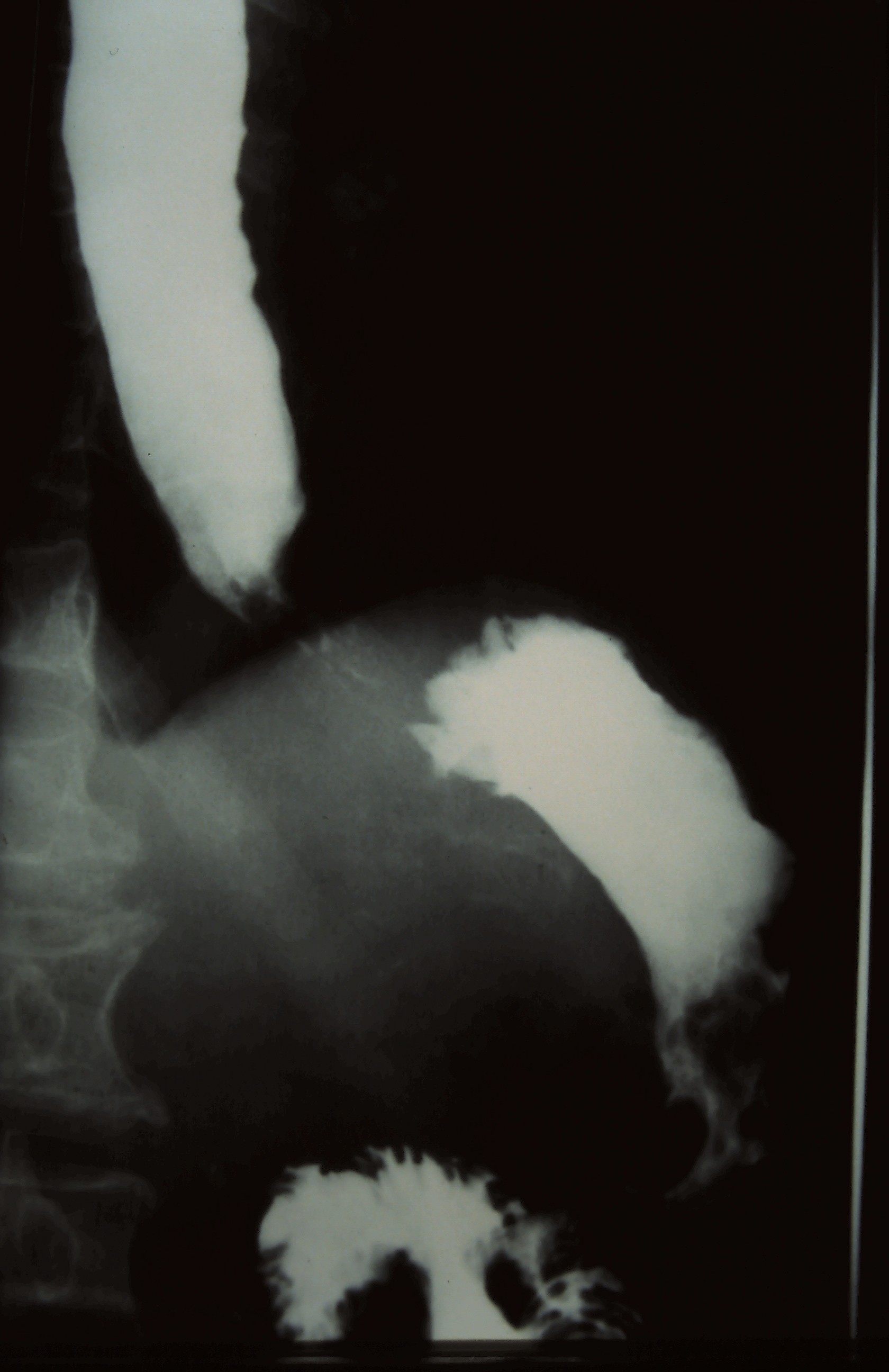
Upper GI: barium-filled stomach.
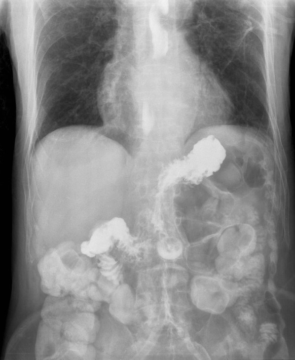
Barium swallow showing reflux.

Quick check

1. An upper GI series is requested for the patient whose erect PA chest radiograph is shown. Based on the findings, which contrast agent is indicated?
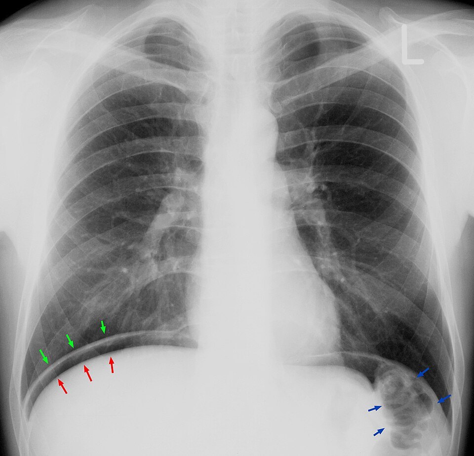

Image: Hellerhoff. CC BY-SA 3.0.

2. During a double-contrast barium enema, an AP right lateral decubitus projection is taken with a horizontal beam. Which parts of the colon are BEST shown with air contrast?

3. A radiographer assisting tableside during a fluoroscopic esophagram wears a wraparound lead apron and a thyroid shield. Why is the thyroid shield recommended in addition to the apron?

4. During a pediatric upper GI, the radiologist reduces the pulsed fluoroscopy rate from 15 to 7.5 pulses per second. What trade-off should the radiographer expect?

5. What fluoroscopic exam is shown in this image?
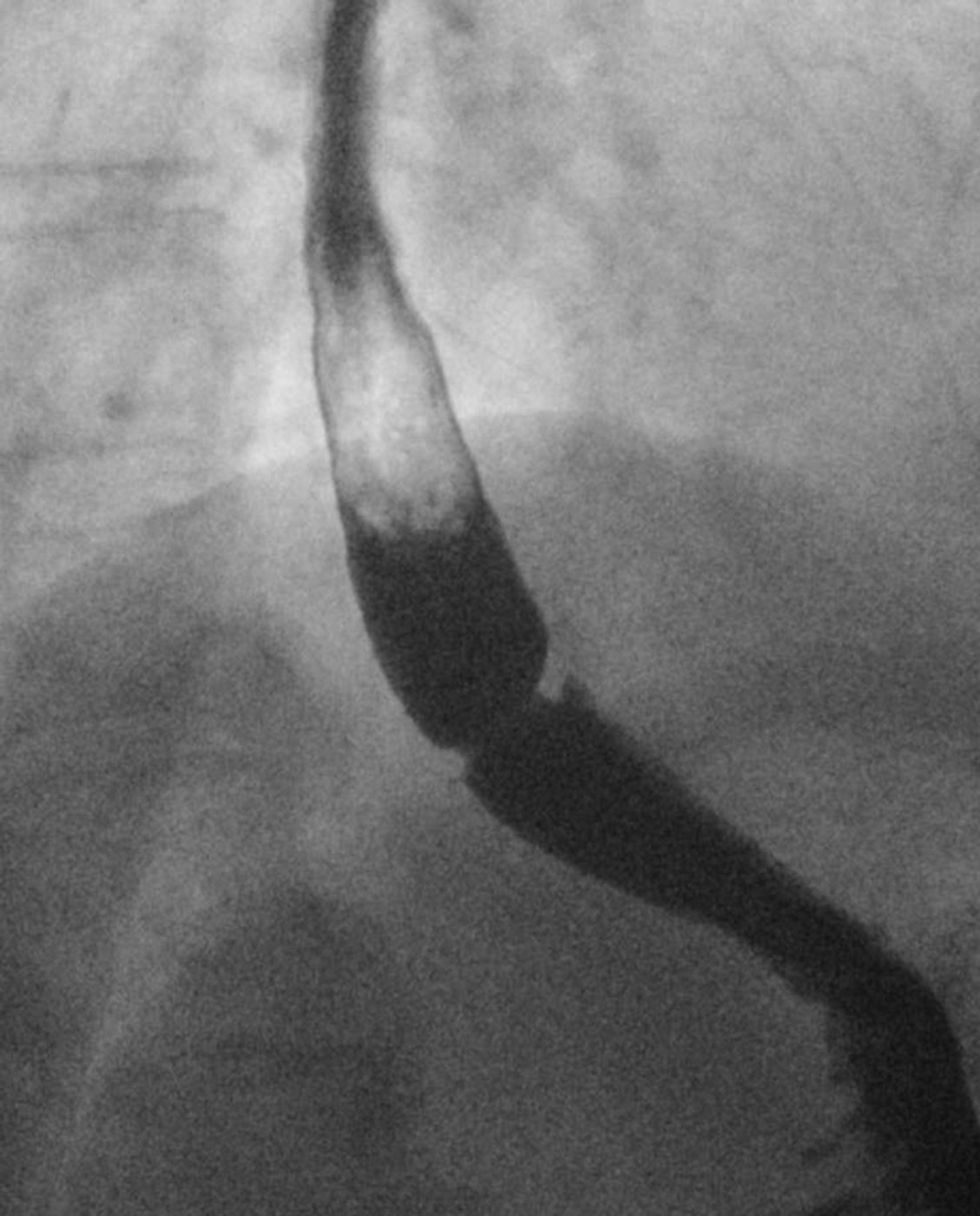
Image: Hellerhoff. CC BY-SA 4.0.

Practice it in 3D

More in the guide