RAD Registry Prep logo

Fluoro & OR Guide

Barium enema

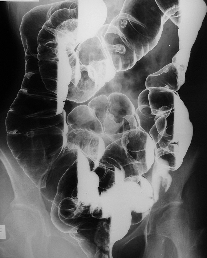
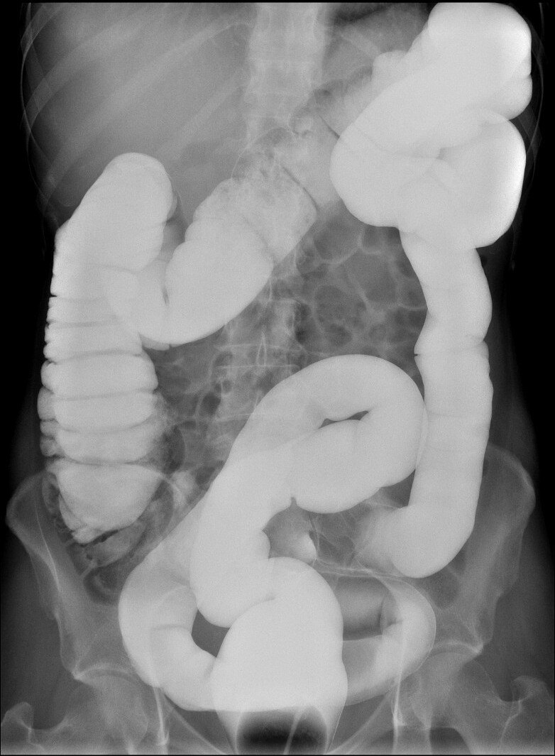
The barium enema (BE, lower GI series) fills the large intestine through a rectal enema tip. A single-contrast BE uses thin barium; a double-contrast (air-contrast) BE uses thick barium and air to coat the lining. You insert the tip, help the patient turn, and take the overhead images.

What the ARRT tests

  • Prep: clear liquid diet and a bowel-cleansing laxative the day before; a clean colon is needed for an accurate exam.
  • Contraindications: suspected perforation (use water-soluble contrast), toxic megacolon, and a colon biopsy in the days before (check your protocol).
  • Tip insertion: patient in the Sims position (left side, right knee flexed). Point the tip toward the umbilicus for the first 1–1.5 inches (2.5–4 cm), then superiorly, never forcing it.
  • Enema bag no more than 24 inches (60 cm) above the table.
  • Obliques: RAO and LPO open the hepatic flexure; LAO and RPO open the splenic flexure.
  • Sigmoid: AP axial (30–40° cephalad) or PA axial (30–40° caudad), also called the butterfly views.
  • Double contrast: right and left lateral decubitus (horizontal beam) show the air-filled sides of the colon; the ventral decubitus lateral shows the rectum.
  • Post-evacuation: a PA or AP image after the patient empties the bowel.

Why it's done

  • Change in bowel habits, rectal bleeding, suspected polyps or tumor, diverticular disease, or inflammatory bowel disease.
  • Water-soluble enemas: leak checks after colon surgery, and some cases of obstruction.
  • Pediatrics: intussusception reduction (with air or contrast) and Hirschsprung disease.
  • Most colon screening is now done with colonoscopy or CT colonography, but the BE is still tested.

Who's in the room

Radiologist
Runs the fluoro, turns the patient for the spot images, and decides on contrast and air.
Radiographer (you)
Prepares the barium and tip, takes the history, inserts the tip, controls flow and air as directed, and takes overheads.

Step by step

  1. Before. Confirm prep, allergies (including latex, for the tip and balloon), pregnancy status and history. Explain the exam and that cramping is normal. Take a scout to check the prep.
  2. Tip. Patient in Sims position. Lubricate the tip, ask the patient to breathe out, and insert it gently as above. Inflate a retention balloon only with the radiologist's approval.
  3. Filling. Under fluoro the barium (and air for a double contrast) fills the colon while the patient turns. The radiologist takes spots of each segment.
  4. Overheads. Your protocol's images, for example PA/AP, RAO, LAO, lateral rectum, decubitus views and an axial sigmoid view.
  5. After. Lower the bag to drain, remove the tip, and help the patient to the restroom. Take the post-evacuation image. Tell the patient to drink plenty of fluids.

Your job

  • Check the scout before starting; a poor prep may cancel the exam.
  • Insert the tip safely and keep the patient covered and informed.
  • Control the flow and air when told; clamp the tubing when the radiologist says stop.
  • Help the patient turn on a narrow table; keep the tubing from pulling on the tip.

Watch out for

  • Perforation: sudden pain, or contrast outside the bowel on fluoro. Stop flow and tell the radiologist.
  • Vasovagal reaction: dizziness, sweating or fainting. Stop, lower the head, and call for help.
  • Elderly or weak patients can fall getting up from the table or in the restroom; stay nearby.
  • Never force the tip; stop if there is resistance or pain.

Tips from the room

  • Tell the patient to take slow, deep breaths through the mouth to ease cramping and help hold the enema.

Images

Double-contrast barium enema: barium coats the colon, air fills it.
Single-contrast barium enema.

Quick check

1. The scout radiograph shown was taken before a scheduled double-contrast barium enema. What is the MOST appropriate action?
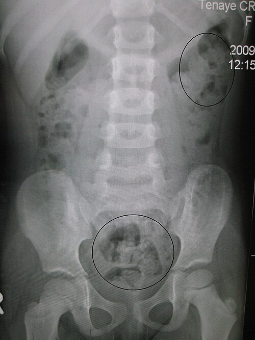

Image: James Heilman, MD. CC BY 3.0.

2. The barium enema shown began with insertion of the rectal enema tip. In which position should the patient be placed for tip insertion?
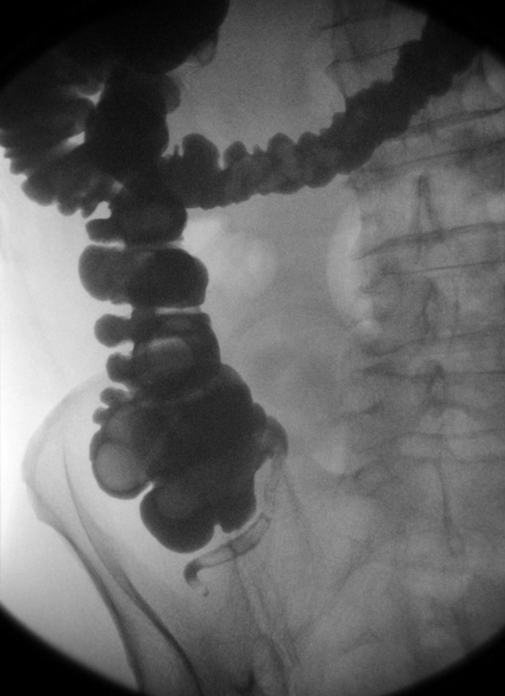
Image: Hellerhoff. CC BY-SA 3.0.

3. During a fluoroscopic barium enema using an image intensifier, the radiologist changes from the 30-cm (12-inch) field to the 15-cm (6-inch) magnification mode to examine a filling defect. How does this change affect the patient's entrance skin dose rate?

4. For a single-contrast barium enema, the radiographer uses 110 to 120 kVp. Why does the image still show high contrast between the barium-filled colon and soft tissue?

5. A patient is scheduled on the same day for a lumbar spine series, a barium enema and an upper GI series. In what order should these exams be performed?

Practice it in 3D

More in the guide