1. This lateral fluoroscopic image is from a swallowing dysfunction study performed with a speech-language pathologist. Why is the lateral projection the primary view for this study?
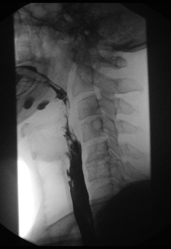

The modified barium swallow (MBS), also called a videofluoroscopic swallow study (VFSS), watches the act of swallowing itself to see whether food or liquid goes into the airway. A speech-language pathologist (SLP) runs the study with the radiologist or radiologist assistant; you set up, run or record the fluoro, and position the patient.
1. This lateral fluoroscopic image is from a swallowing dysfunction study performed with a speech-language pathologist. Why is the lateral projection the primary view for this study?
2. During a modified barium swallow, which team member typically selects the bolus consistencies and tests swallowing strategies while the radiographer positions the patient and records the study?
3. During a modified barium swallow, which bolus is used to evaluate chewing and the oral preparation of a solid?
4. Why is a modified barium swallow recorded as continuous fluoroscopic video rather than documented with last-image-hold or spot images?
5. A patient is referred for a modified barium swallow rather than a routine esophagram. Which phases of swallowing are the main focus of the modified barium swallow?
The parts, the movements, the dose modes, and where to stand.
What's sterile, what you can touch, and how to move around the field.
Barium swallow and UGI: why they're ordered, the positions, and your role.
Timed films as barium moves from the stomach to the colon.
Single- and double-contrast BE: prep, tip insertion, positions, and overheads.
Bladder filled through a catheter: leak checks, reflux, and voiding images.
Uterus and fallopian tubes for infertility: timing, setup, and dose.
Joint injection under fluoro, usually before MRI or CT.
Contrast in the spinal canal: needle site, table tilt, and keeping contrast out of the head.
Spinal tap under fluoro when bedside attempts fail.
Bile ducts through a scope, in surgery, or through a T-tube.
The most common C-arm case: the fracture table, the two views, and your role.
Open reduction and internal fixation of the ankle or wrist: the views, the mini C-arm, and hands in the beam.