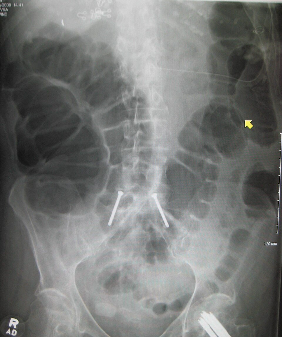
1. Which feature identifies the dilated loops on this abdominal radiograph as large bowel rather than small bowel?

The small bowel series (small bowel follow-through, SBFT) follows barium from the stomach through the jejunum and ileum to the large bowel. It's often added to the end of an upper GI. Most of it is timed overhead images that you take; the radiologist adds fluoro spots of the terminal ileum at the end.
1. Which feature identifies the dilated loops on this abdominal radiograph as large bowel rather than small bowel?
2. During a small bowel follow-through, the terminal ileum shows a pattern of crisscrossing ulcers with raised islands of swollen mucosa between them, giving a 'cobblestone' appearance. Which condition is MOST likely?
3. During a routine small bowel follow-through, when are overhead images typically taken?
4. In a double-contrast enteroclysis, what is infused after the barium to distend the small bowel?
5. During ileocecal spot imaging in a small bowel follow-through, why does the radiologist apply a compression paddle over the right lower quadrant?
The parts, the movements, the dose modes, and where to stand.
What's sterile, what you can touch, and how to move around the field.
Barium swallow and UGI: why they're ordered, the positions, and your role.
Video swallow study with speech therapy: bolus types, the field of view, and your role.
Single- and double-contrast BE: prep, tip insertion, positions, and overheads.
Bladder filled through a catheter: leak checks, reflux, and voiding images.
Uterus and fallopian tubes for infertility: timing, setup, and dose.
Joint injection under fluoro, usually before MRI or CT.
Contrast in the spinal canal: needle site, table tilt, and keeping contrast out of the head.
Spinal tap under fluoro when bedside attempts fail.
Bile ducts through a scope, in surgery, or through a T-tube.
The most common C-arm case: the fracture table, the two views, and your role.
Open reduction and internal fixation of the ankle or wrist: the views, the mini C-arm, and hands in the beam.