
1. A patient with a suspected herniated nucleus pulposus is scheduled for lumbar myelography. Into which space is the contrast medium injected?
Lumbar puncture (fluoro-guided)
A lumbar puncture (LP, spinal tap) places a needle into the spinal canal to take a sample of cerebrospinal fluid (CSF), measure pressure, or give medicine. Many are done at the bedside; patients come to fluoro when that fails or when the spine is hard to reach.
What the ARRT tests
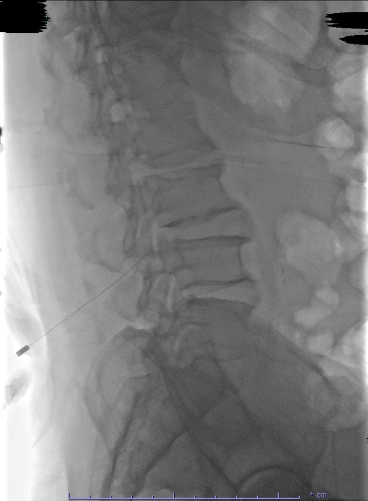
- Needle level: L3–L4 or L4–L5 most often (below the end of the spinal cord at about L1–L2).
- Patient prone with a pillow under the abdomen, or in a lateral decubitus position if opening pressure is needed.
- CSF tubes are numbered and filled in order, then sent with their labels.
- Contraindications: increased intracranial pressure from a mass (risk of brain herniation), a bleeding problem or blood thinners, and infection at the site.
- Opening pressure is measured with a manometer, ideally with the patient lying on their side and relaxed.
Why it's done
- Suspected meningitis, bleeding around the brain, or other neurological conditions.
- Measuring CSF pressure, or removing fluid to lower it.
- Giving chemotherapy or other medicine into the CSF.
- Referred to fluoro after failed bedside attempts, obesity, scoliosis, or spine surgery.
Who's in the room
- Radiologist or radiologist assistant
- Places the needle under fluoro, collects the fluid, and measures pressure.
- Radiographer (you)
- Sets up the tray, positions the patient, runs the fluoro, and labels and sends the specimens.
Step by step
- Before. Confirm consent, blood thinners and lab values per protocol, allergies, and whether opening pressure is needed. Set up the LP kit and numbered tubes.
- Needle. Patient prone with a pillow under the abdomen. The radiologist numbs the skin and advances the needle under fluoro.
- Fluid. CSF is collected into tubes 1 to 4 in order. Pressure is measured if ordered.
- After. Bandage the site, label and send the tubes right away, and explain aftercare: rest and fluids, and report a severe headache, fever, or leg weakness.
Your job
- Specimen handling: labels on the tubes, in order, sent right away (some tests need to be run fast).
- Pulsed fluoro with last-image hold; the needle is placed mostly from saved images.
Watch out for
- Headache after an LP is common; worse when upright, better lying down.
- Back pain with new leg weakness or numbness may mean bleeding in the spinal canal: report at once.
Images
Quick check
2. A patient arrives for a lumbar myelogram. At the planned puncture site, the radiographer notices a pustule with surrounding redness. What is the MOST appropriate action?
3. Which task is within the radiographer's role during a fluoroscopically guided lumbar puncture for myelography?
4. A spinal needle used for a lumbar puncture contains a removable stylet. What is the main purpose of the stylet?
5. The needle tip in this fluoroscopy-guided lumbar puncture lies between L3 and L4. Why is an adult lumbar puncture performed at L3-L4 or L4-L5?
Practice it in 3D
More in the guide
C-arm basics
The parts, the movements, the dose modes, and where to stand.
Sterile basics
What's sterile, what you can touch, and how to move around the field.
Esophagram and upper GI series
Barium swallow and UGI: why they're ordered, the positions, and your role.
Modified barium swallow (swallow study)
Video swallow study with speech therapy: bolus types, the field of view, and your role.
Small bowel follow-through
Timed films as barium moves from the stomach to the colon.
Barium enema
Single- and double-contrast BE: prep, tip insertion, positions, and overheads.
Cystogram and VCUG
Bladder filled through a catheter: leak checks, reflux, and voiding images.
Hysterosalpingogram (HSG)
Uterus and fallopian tubes for infertility: timing, setup, and dose.
Arthrogram
Joint injection under fluoro, usually before MRI or CT.
Myelogram
Contrast in the spinal canal: needle site, table tilt, and keeping contrast out of the head.
ERCP and cholangiograms
Bile ducts through a scope, in surgery, or through a T-tube.
Hip pinning (hip fracture fixation)
The most common C-arm case: the fracture table, the two views, and your role.
Ankle and wrist ORIF
Open reduction and internal fixation of the ankle or wrist: the views, the mini C-arm, and hands in the beam.