The wrist is eight carpal bones in two rows, sitting on the distal radius and ulna. Routine wrist imaging is a PA, a PA oblique and a lateral, all centered to the midcarpal area, with special views for the scaphoid and the carpal canal.
Overview
Bones
Proximal row (thumb side to little-finger side)
Scaphoid
Lunate
Triquetrum
Pisiform
Distal row (thumb side to little-finger side)
Trapezium
Trapezoid
Capitate
Hamate
Forearm
Distal radius (thumb side)
Distal ulna (little-finger side)
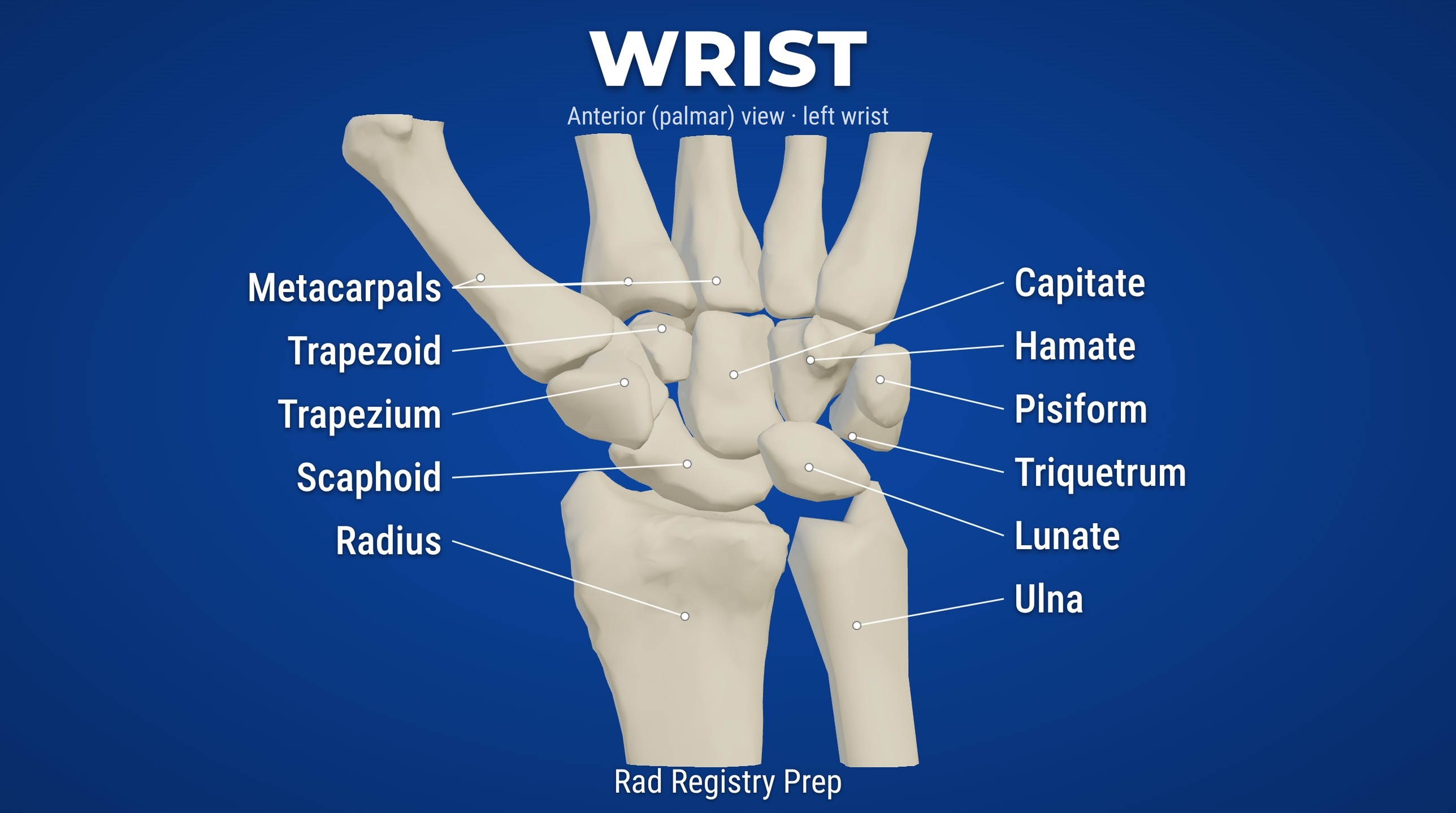
Some Lovers Try Positions That They Can't Handle. Read each row from the thumb side: S-L-T-P across the proximal row, then T-T-C-H across the distal row. The thumb always sits on the trapezium.
Registry must-knows
The scaphoid is the most commonly fractured carpal bone. Snuffbox tenderness calls for a PA in ulnar deviation, CR perpendicular or angled 10–15° toward the elbow.
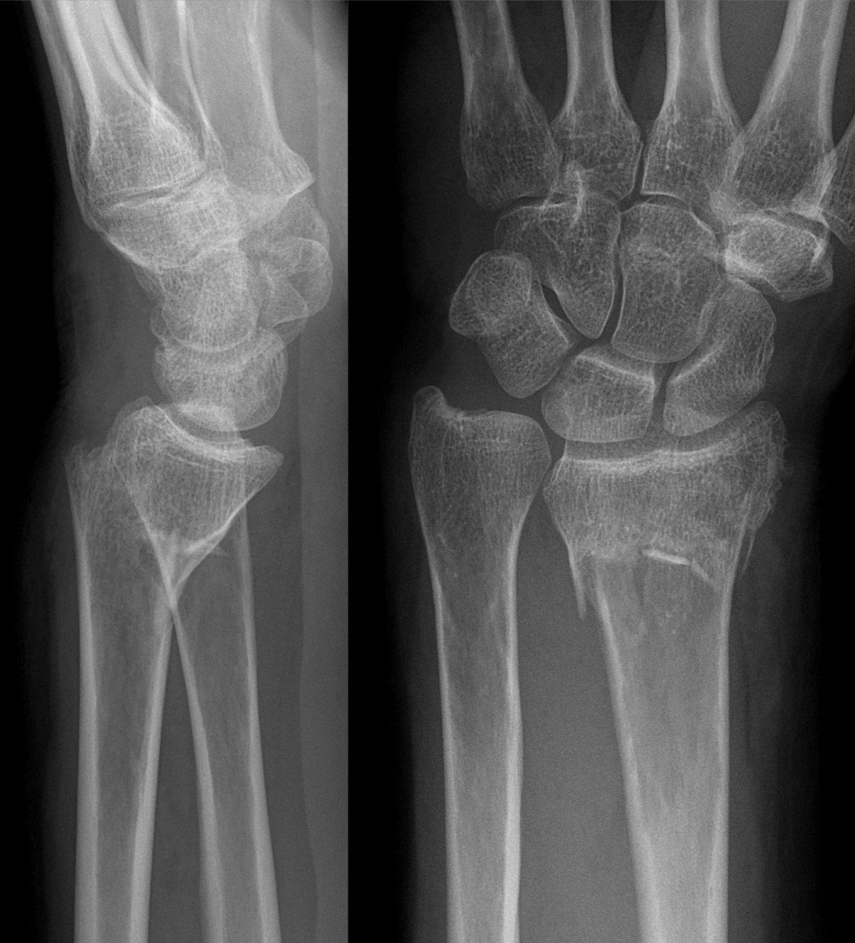
Colles fracture: distal radius with the fragment displaced posteriorly (dorsal). Smith fracture: displaced anteriorly (volar). Displacement is judged on a true lateral.
On a true lateral, the radius, lunate, capitate and 3rd metacarpal line up and the distal radius and ulna are superimposed. Lunate and perilunate dislocations break that line.
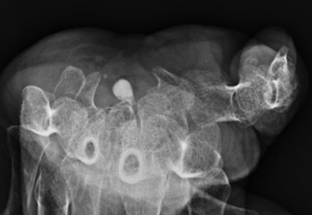
The Gaynor-Hart (tangential) projection shows the carpal canal, the pisiform and the hook of the hamate.
Show 1 more
Flexing the fingers slightly on the PA brings the carpals into contact with the IR and reduces OID.
3D model
Loading 3D…
Radiographs 6
Pick a film, then a structure to circle it.
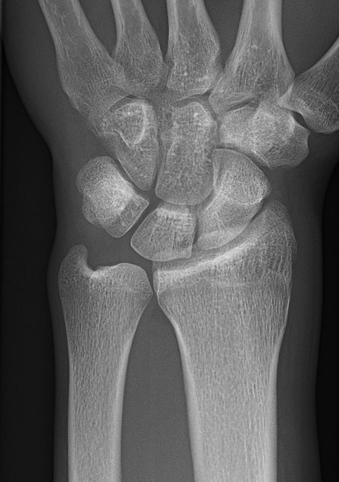
PA wristImage: Mikael Häggström, M.D., via Wikimedia Commons. CC0 (public domain dedication). Source
Elbow flexed 90°, hand pronated, fingers slightly flexed so the carpals rest on the IR.
Central ray
Perpendicular
Centering
Midcarpal area
Typical technique
55–60 kVp · 2–3 mAs · 40" SID
Best shows
Carpals, distal radius and ulna, proximal metacarpals
Image criteria
Distal radius and ulna, carpals and proximal metacarpals included.
No rotation: radius and ulna side by side with only slight overlap at the distal radioulnar joint.
Some carpals overlap normally; the scaphoid looks foreshortened.
PA oblique wrist (lateral rotation)
CR: Perpendicular
Position
From the PA, rotate the hand and wrist 45° laterally (thumb side up), supported on a sponge.
Central ray
Perpendicular
Centering
Midcarpal area
Typical technique
55–60 kVp · 2–3 mAs · 40" SID
Best shows
The lateral carpals: trapezium and scaphoid, with less overlap than on the PA.
Image criteria
Trapezium and scaphoid well seen with minimal superimposition.
Slight overlap of the distal radius and ulna; the ulnar carpals partly overlap.
Carpals, distal radius and ulna and proximal metacarpals included.
Lateral wrist (lateromedial)
CR: Perpendicular
Position
Elbow flexed 90°, ulnar side of the wrist on the IR, hand and wrist in a true lateral.
Central ray
Perpendicular
Centering
Wrist joint (midcarpal area)
Typical technique
60–65 kVp · 3–4 mAs · 40" SID
Best shows
Anterior or posterior displacement of fractures (e.g., Colles, Smith) and the lunate–capitate alignment.
Image criteria
Distal radius and ulna superimposed (ulnar head over the radius).
Metacarpals superimposed; the thumb may sit slightly anterior.
No rotation: radius, lunate, capitate and 3rd metacarpal line up.
PA in ulnar deviation (scaphoid view)
CR: Perpendicular, or angled 10–15° proximally (toward the elbow)
Position
Hand pronated as for the PA, then turned toward the ulnar side without moving the forearm.
Central ray
Perpendicular, or angled 10–15° proximally (toward the elbow)
Centering
Scaphoid (anatomic snuffbox)
Typical technique
55–60 kVp · 2–3 mAs · 40" SID
Best shows
The scaphoid in its full length, so a waist fracture can be seen
Image criteria
Scaphoid shown in its full length without foreshortening.
Open spaces around the scaphoid, especially on its lateral side.
Wrist in ulnar deviation with no rotation of the forearm.
Scaphoid PA axial
StecherCR: Perpendicular to the tabletop, or angled 20° toward the elbow if the IR is flat
Position
PA wrist with the IR (or the fist) elevated 20°.
Central ray
Perpendicular to the tabletop, or angled 20° toward the elbow if the IR is flat
Centering
Scaphoid
Typical technique
55–60 kVp · 2–3 mAs · 40" SID
Best shows
The scaphoid elongated, free of superimposition
Image criteria
Scaphoid shown without foreshortening.
Spaces around the scaphoid open.
Carpal canal (tangential)
Gaynor-HartCR: 25–30° to the long axis of the hand
Position
Hyperextend the wrist (patient pulls the fingers back) and rotate the hand about 10° internally.
Central ray
25–30° to the long axis of the hand
Centering
About 1" distal to the base of the 3rd metacarpal
Typical technique
60–65 kVp · 3–5 mAs · 40" SID
Best shows
The carpal canal, pisiform and hook of the hamate
Image criteria
Carpal canal shown in profile, rounded and open.
Pisiform and hook of the hamate separated.
Technique values are typical starting points; follow your department's technique chart.
Labeled diagrams
Wrist (anterior (palmar), left wrist view) · Diagram: Rad Registry Prep (original illustration).
Quick check
1. On the lateral projection of this wrist pair, the extra-articular distal radius fracture shows the distal fragment displaced anteriorly (volar). This fracture is classified as a:
Image: Hellerhoff. CC BY-SA 4.0.
2. On a PA wrist of an 8-year-old, a smooth, regular lucent line crosses the distal radius at the junction of the epiphysis and metaphysis, and a matching line is present on the distal ulna. What does this MOST likely represent?
3. Which wrist projection BEST demonstrates the trapezium and scaphoid with the least superimposition?
4. While reviewing a PA wrist image, a radiographer zooms in to 4× on the scaphoid to check for a fracture line. How does zooming affect the image's spatial resolution?
5. This tangential view looks down the carpal tunnel and shows a round calcification inside it. How is this carpal canal (Gaynor-Hart) projection positioned?
Image: Woo SH, Choi BG, Woo SJ, Park KH, Arch Plast Surg 2026, via PubMed Central. CC BY 4.0.