
1. A femoral shaft fracture appears well aligned on the AP projection. What is the MOST important reason a lateral projection is still required?
Femur
The femur is the longest and strongest bone. Routine femur imaging is an AP and a lateral; most adults need two images per view so the hip and the knee are both included.
Overview
Bones
Proximal
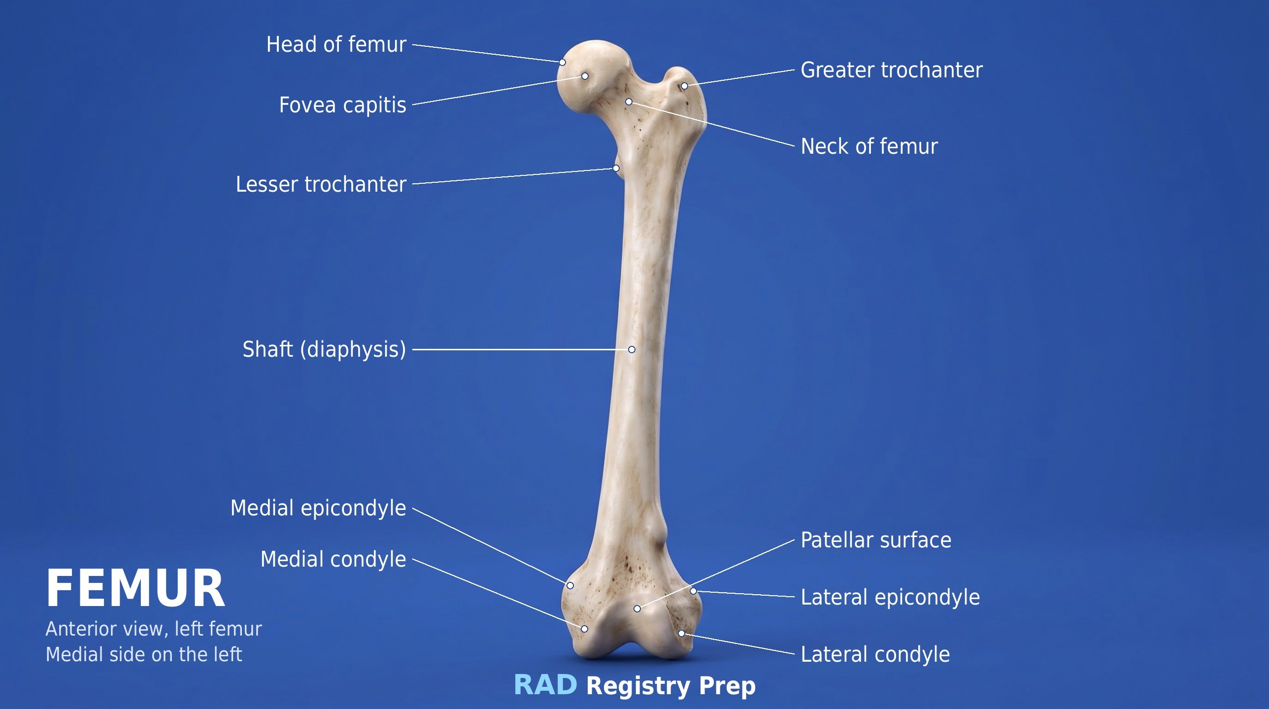
- Head
- Neck
- Greater trochanter
- Lesser trochanter
Shaft
- Linea aspera (posterior)
Distal
- Medial condyle
- Lateral condyle
- Medial epicondyle
- Lateral epicondyle
- Adductor tubercle
- Intercondylar fossa
- Patellar surface
Registry must-knows
- Include both joints: often two exposures, one with the hip and one with the knee, overlapping in the middle.
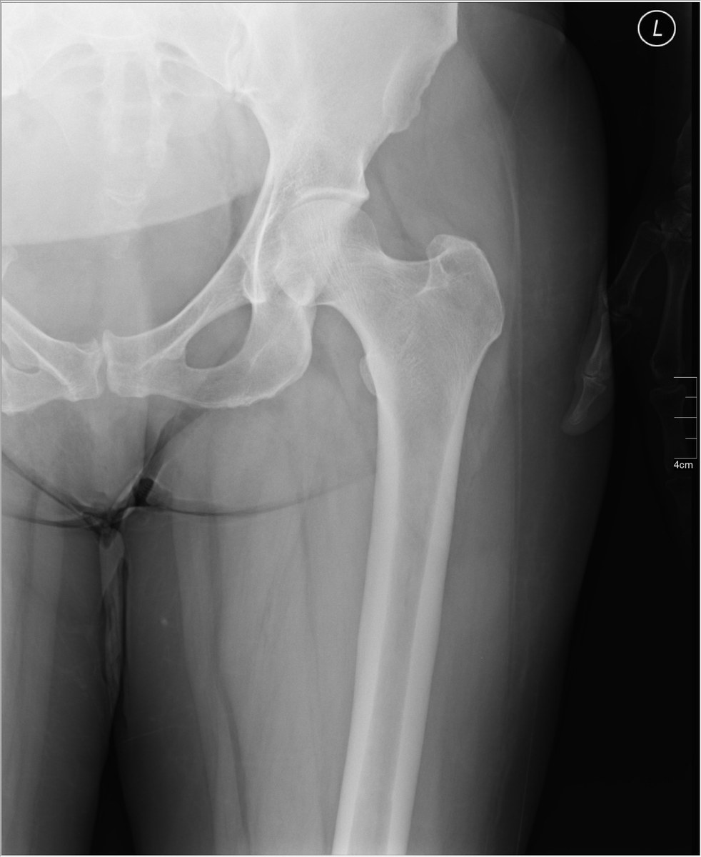
- AP: rotate the leg 15–20° internally (unless a fracture is suspected) so the femoral neck is in profile.
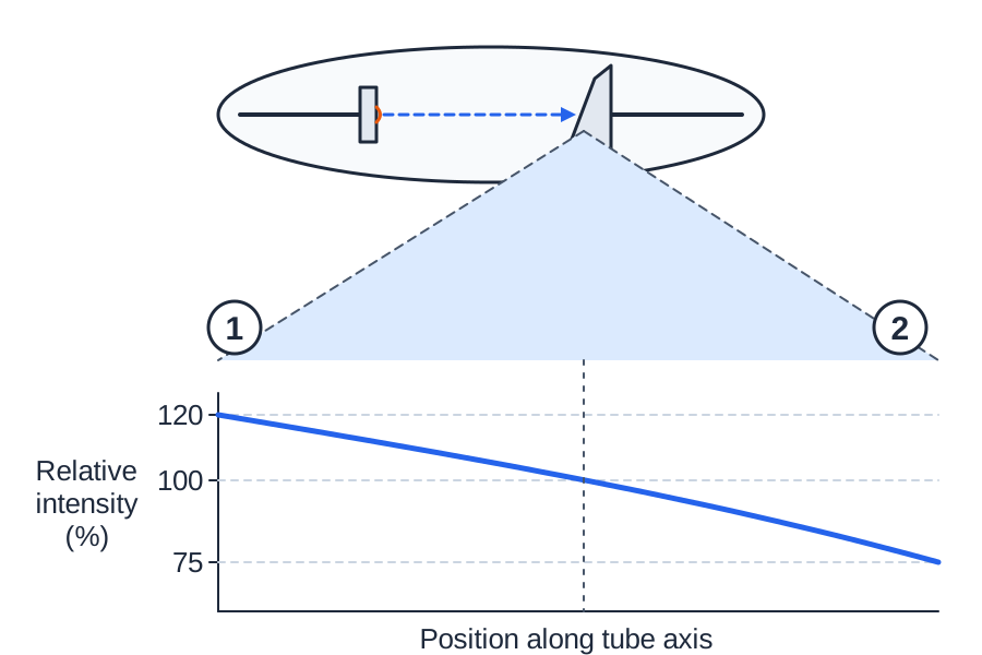
- Use the anode heel effect: put the cathode over the thicker hip end.
- Never frog-leg a suspected proximal femur fracture; use a cross-table lateral.
Show 1 more
- The adductor tubercle on the medial condyle helps tell a true lateral of the distal femur: the condyles should superimpose.
3D model
Loading 3D…
Radiographs 2
Pick a film, then a structure to circle it.
Projections 2
Tap a projection for positioning, central ray and image criteria. See every exam in the Positioning Guide ›
AP femur
CR: Perpendicular
- Position
- Supine, leg rotated 15–20° internally (not in trauma); one image with the hip and one with the knee if needed.
- Central ray
- Perpendicular
- Centering
- Mid-femur (or the center of each part)
- Typical technique
- 75–80 kVp · 10–20 mAs · 40" SID
Image criteria
- Hip and knee joints both included (on one or two images).
- Femoral neck not foreshortened; lesser trochanter barely seen.
- Femoral and tibial condyles symmetric at the knee.
Lateral femur
CR: Perpendicular
- Position
- Lateral recumbent on the affected side, knee flexed about 45°, opposite leg behind. Trauma: cross-table lateral.
- Central ray
- Perpendicular
- Centering
- Mid-femur
- Typical technique
- 75–80 kVp · 10–20 mAs · 40" SID
Image criteria
- Femoral condyles superimposed at the distal end.
- Patella in profile; patellofemoral space open.
- Hip and knee both included (one or two images).
Technique values are typical starting points; follow your department's technique chart.
Labeled diagrams
Quick check
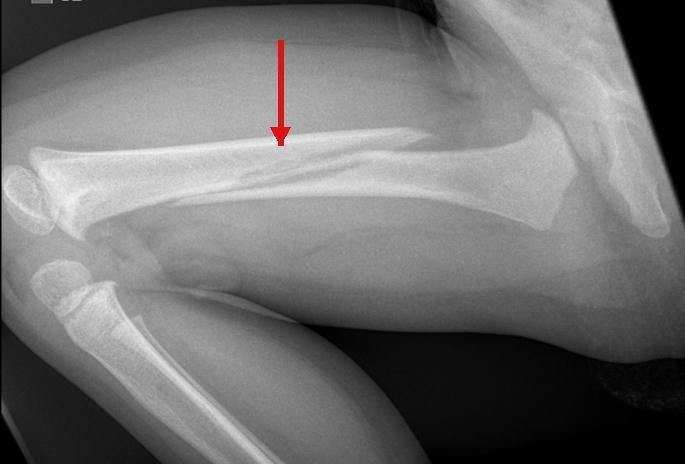
2. A 4-month-old infant who is not yet crawling has the femoral shaft fracture indicated by the red arrow, and the history does not explain the injury. The radiologist requests imaging to evaluate for non-accidental trauma. Which examination should the radiographer perform?
3. The diagram shows an x-ray tube (cathode filament on the left, angled anode target on the right) above a graph of relative beam intensity along the tube axis. For an AP femur on a large adult, where should the hip be placed to use the anode heel effect?
4. An elderly patient arrives from the ED after a fall. The left leg is shortened and the foot is turned outward, and a hip fracture is suspected. How should the radiographer position the leg for the AP femur and hip?
5. On this AP of the proximal femur, the lesser trochanter is barely seen. How was the leg positioned?