The pelvis is the two hip bones (each an ilium, ischium and pubis fused at the acetabulum), the sacrum and the coccyx. The routine AP pelvis turns the feet in 15–20° so the femoral necks show in full length.
Overview
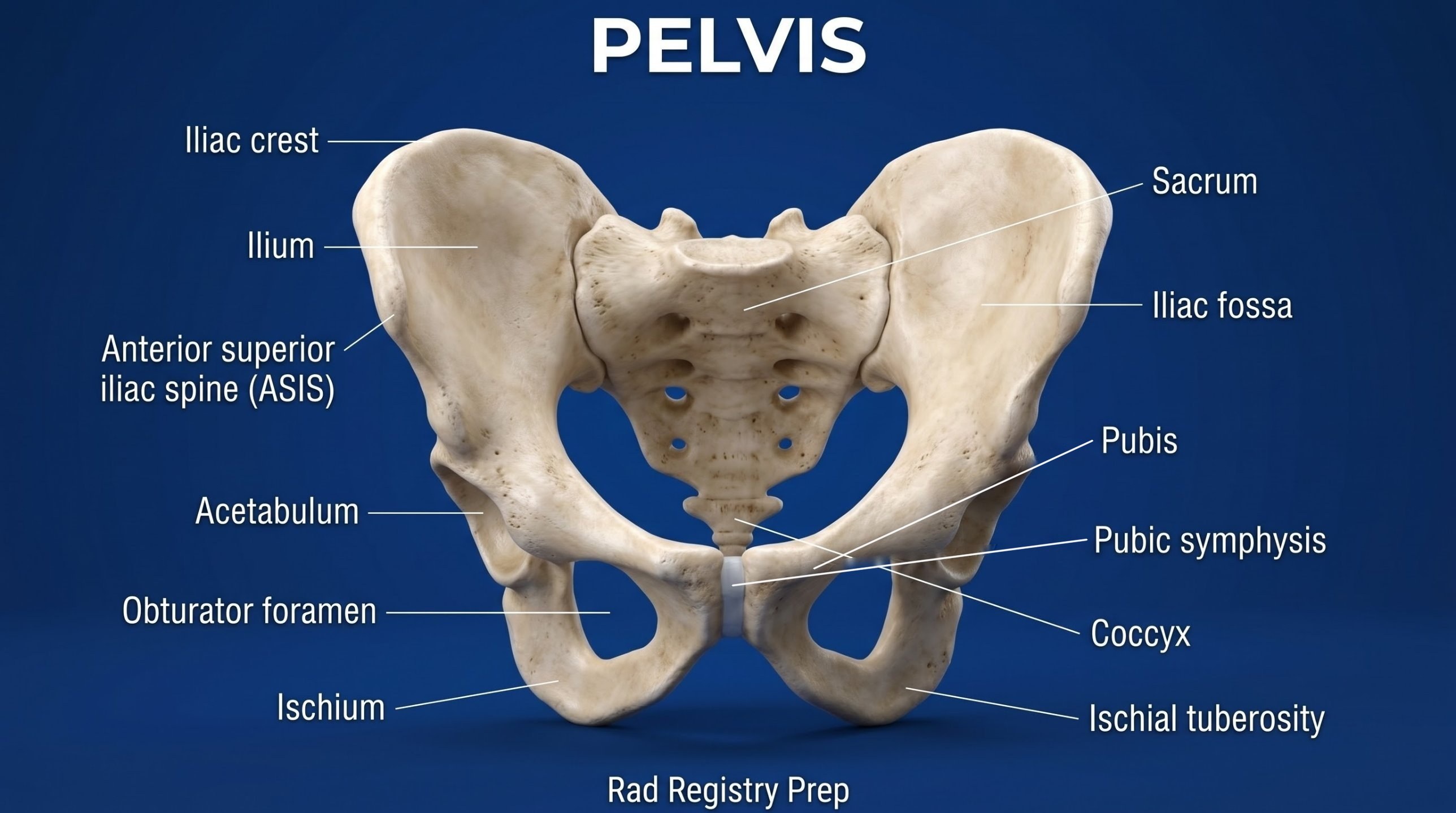
Bones
Hip bone (os coxae)
Ilium
Ischium
Pubis
Acetabulum
Landmarks
Iliac crest
ASIS
Ischial tuberosity
Pubic symphysis
Obturator foramen
Spine
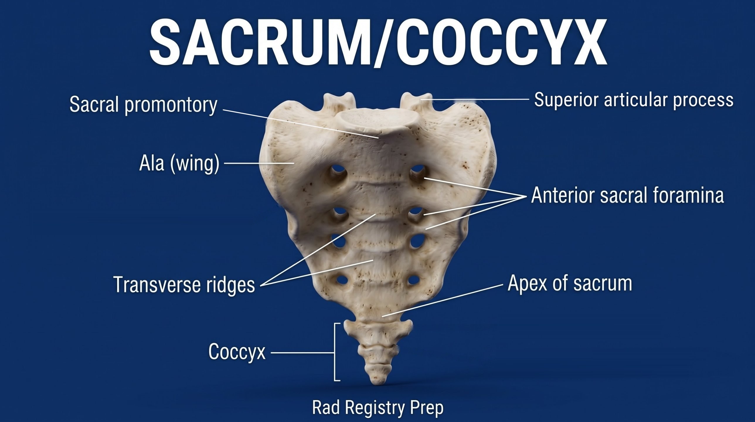
Sacrum
Sacral promontory
Coccyx
SI joints
The iliac crest is level with L4–L5; the ASIS is level with S1–S2; the greater trochanter is level with the pubic symphysis.
Registry must-knows
AP pelvis: rotate the feet and legs 15–20° internally to lengthen the femoral necks, unless a hip fracture is suspected.
With the legs rotated in, the lesser trochanters are barely seen; if they're prominent, the legs are externally rotated.
A female pelvis is wider and shallower, with a larger, rounder inlet and a pubic arch angle over 90°.
Inlet view: CR 40° caudad. Outlet view (Taylor): CR 20–35° cephalad for men, 30–45° for women.
Show 1 more
SI joints AP axial: CR 30–35° cephalad, entering 2 inches below the ASIS.
3D model
Loading 3D…
Radiographs 11
Pick a film, then a structure to circle it.
AP pelvisImage: UC San Diego Health / bonepit.com. Public domain. Source
Supine, MSP centered, legs extended with the feet and legs rotated 15–20° internally (not in trauma).
Central ray
Perpendicular
Centering
Midway between the ASIS level and the pubic symphysis (about 2 inches below the ASIS)
Typical technique
80–85 kVp · 20–40 mAs · 40" SID
Image criteria
Entire pelvis and the proximal femurs included.
No rotation: symmetric iliac wings and obturator foramina.
Femoral necks in full length; greater trochanters in profile, lesser trochanters barely seen.
AP axial inlet
CR: 40° caudad
Position
Supine, as for the AP pelvis.
Central ray
40° caudad
Centering
At the level of the ASIS, on the MSP
Typical technique
80–90 kVp · 20–40 mAs · 40" SID
Best shows
The pelvic ring (inlet) in its entirety, for displacement after trauma
Image criteria
Pelvic ring seen as an open ring.
Symmetric, no rotation.
AP axial outlet
TaylorCR: 20–35° cephalad (men), 30–45° (women)
Position
Supine, as for the AP pelvis.
Central ray
20–35° cephalad (men), 30–45° (women)
Centering
1–2 inches below the superior border of the pubic symphysis
Typical technique
80–90 kVp · 20–40 mAs · 40" SID
Best shows
Superior and inferior pubic and ischial rami, with little foreshortening
Image criteria
Pubic and ischial rami elongated.
Obturator foramina symmetric.
AP oblique pelvis, both sides
Judet RPO / LPOCR: Perpendicular
Position
Semi-supine, body rotated 45° (sponge under the raised side), both legs extended. RPO: right side down. LPO: left side down. Both are taken.
Central ray
Perpendicular
Centering
As for the AP pelvis, on a landscape 14 × 17 detector (some departments cone to one acetabulum instead: see Hip)
Typical technique
80–85 kVp · 20–40 mAs · 40" SID
Best shows
Both acetabula on one film: the DOWNSIDE as an iliac oblique (posterior column, anterior rim, iliac wing open), the UPSIDE as an obturator oblique (anterior column, posterior rim, obturator foramen open)
Image criteria
Both iliac crests, acetabula and proximal femurs included.
Downside: iliac wing open and in profile, obturator foramen closed.
Supine, body rotated 25–30° with the side of interest UP: LPO shows the right SI joint, RPO the left.
Central ray
Perpendicular
Centering
1 inch medial to the elevated ASIS
Typical technique
80–85 kVp · 20–30 mAs · 40" SID
Best shows
The SI joint farther from the IR, open
Image criteria
Joint space open, with no overlap of the ilium and sacrum.
Both sides done for comparison.
AP axial sacrum
CR: 15° cephalad
Position
Supine, MSP centered; empty bladder and colon if possible.
Central ray
15° cephalad
Centering
2 inches superior to the pubic symphysis
Typical technique
80–85 kVp · 20–30 mAs · 40" SID
Image criteria
Sacrum free of foreshortening, sacral foramina seen.
Symphysis pubis not superimposed over the sacrum.
No rotation.
AP axial coccyx
CR: 10° caudad
Position
Supine, MSP centered; empty bladder.
Central ray
10° caudad
Centering
2 inches superior to the pubic symphysis
Typical technique
75–80 kVp · 15–25 mAs · 40" SID
Image criteria
Coccygeal segments not superimposed by the pubis.
Coccyx centered, no rotation.
Lateral sacrum and coccyx
CR: Perpendicular
Position
Lateral recumbent, knees and hips flexed, MSP parallel to the table. Lead mat on the table behind the patient to cut scatter.
Central ray
Perpendicular
Centering
Sacrum: 3–4 inches posterior to the ASIS. Coccyx: 3–4 inches posterior and 2 inches inferior to the ASIS.
Typical technique
85–95 kVp · 30–60 mAs · 40" SID
Image criteria
Sacrum and coccyx in profile; greater sciatic notches and femoral heads superimposed.
L5–S1 included for the sacrum.
Technique values are typical starting points; follow your department's technique chart.
Labeled diagrams
Pelvis (anterior view) · Diagram: Rad Registry Prep (original illustration).Sacrum and Coccyx (anterior view) · Diagram: Rad Registry Prep (original illustration).
Quick check
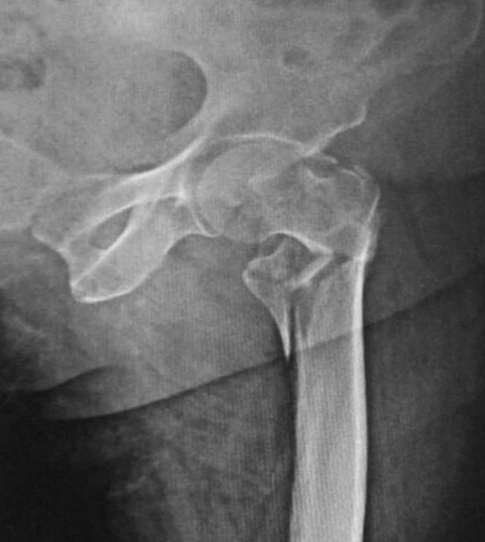
1. This AP hip radiograph of a patient in the emergency department shows a displaced, comminuted proximal femur fracture. The patient can flex and raise the unaffected leg. Which lateral projection should the radiographer perform?
Image: Memon, Patel & Juva. CC BY 4.0.
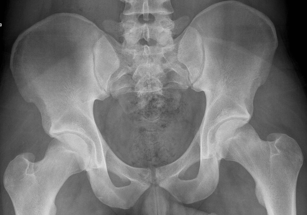
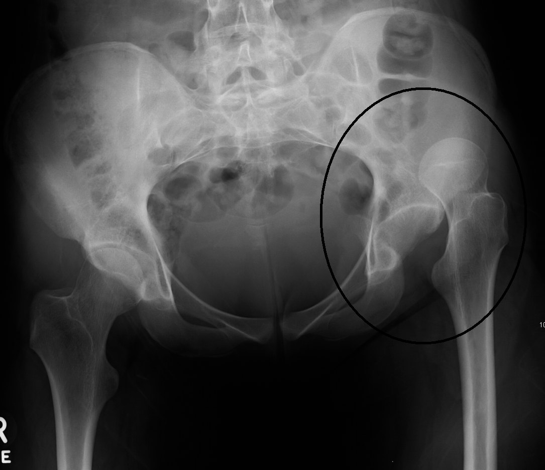
2. This AP pelvis was taken after a car crash in which the patient's knee struck the dashboard. The left femoral head lies superior and lateral to the acetabulum. Which injury is MOST likely?
Image: James Heilman, MD. CC BY-SA 3.0.
3. An AP pelvis on a trauma patient shows a possible acetabular fracture. Which additional projections BEST demonstrate the anterior and posterior columns of the acetabulum?
4. Lumbar spine and pelvis radiographs show fused sacroiliac joints and thin bony bridges joining the vertebral bodies, producing a 'bamboo spine.' Which condition is MOST likely?
5. On a standing AP pelvis, the obturator foramina and iliac wings are symmetric, but the right iliac crest and right femoral head are higher than the left. The patient's weight was equal on both feet. What does this MOST likely indicate?