The foot has 26 bones: 7 tarsals, 5 metatarsals and 14 phalanges. A routine foot is an AP (dorsoplantar), a medial oblique and a lateral.
Overview
Bones
Tarsals (7)
Calcaneus
Talus
Navicular
Cuboid
Medial cuneiform
Intermediate cuneiform
Lateral cuneiform
Metatarsals (5)
1st (great toe)
2nd
3rd
4th
5th
Phalanges (14)
Great toe: proximal and distal
Toes 2–5: proximal, middle and distal
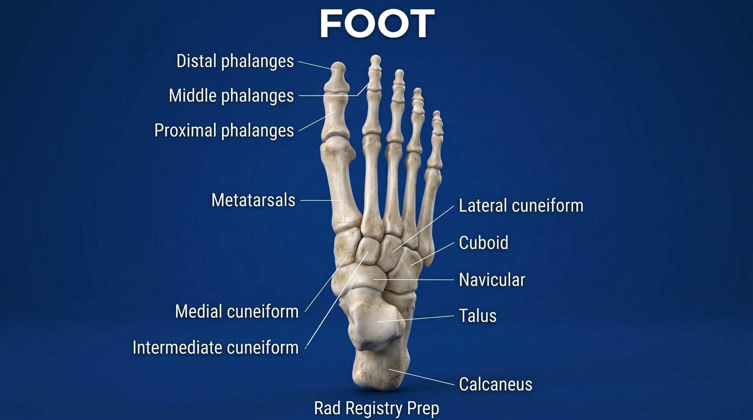
Seven tarsals: calcaneus, talus, navicular, cuboid and three cuneiforms (medial, intermediate, lateral). The talus sits on the calcaneus; the cuboid is lateral, the navicular medial.
Registry must-knows
AP foot: CR 10° toward the heel (perpendicular to the metatarsals), centered to the base of the 3rd metatarsal.
Medial oblique (30–40°) best shows the cuboid, the base of the 5th metatarsal and the sinus tarsi.
Jones fracture: the proximal shaft of the 5th metatarsal; avulsion fractures happen at its tuberosity.
Weight-bearing AP and lateral feet show the longitudinal arches (flat foot).
Show 2 more
Toes: center to the MTP joint of the toe of interest; the great toe has two phalanges, the others have three.
Calcaneus: an axial (plantodorsal, CR 40° cephalad) and a lateral. Falls from a height cause calcaneal fractures; check the spine too.
3D model
Loading 3D…
Radiographs 9
Pick a film, then a structure to circle it.
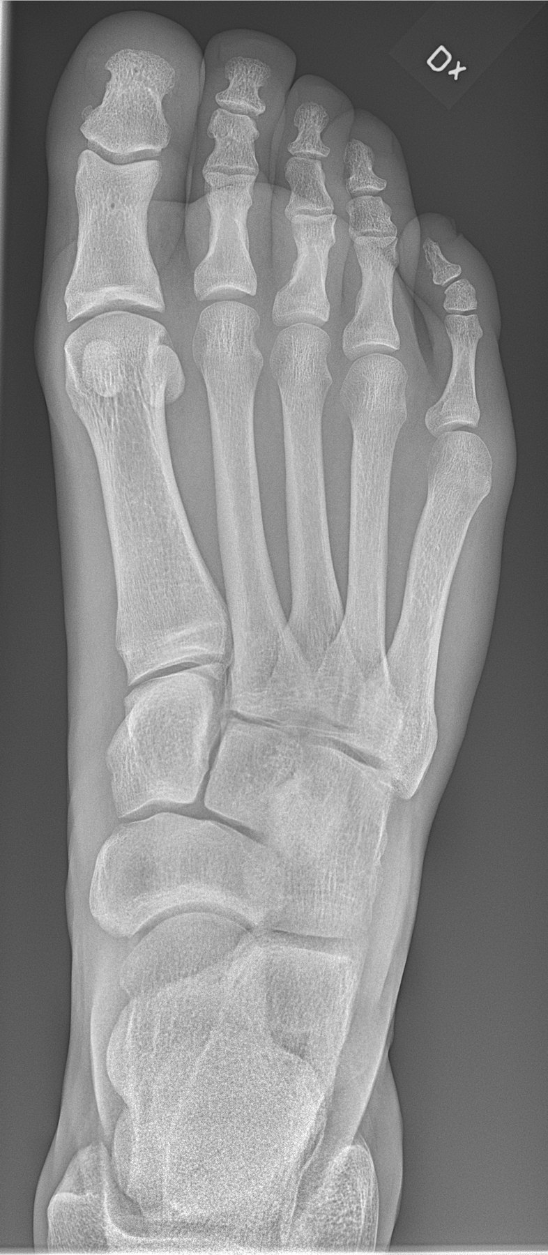
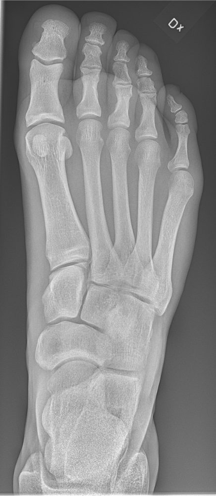
AP footImage: Mikael Häggström, M.D., via Wikimedia Commons. CC0 (public domain dedication). Source
CR: 10° toward the heel (perpendicular to the metatarsals)
Position
Supine or seated, knee flexed, plantar surface flat on the IR.
Central ray
10° toward the heel (perpendicular to the metatarsals)
Centering
Base of the 3rd metatarsal
Typical technique
55–65 kVp · 2–4 mAs · 40" SID
Image criteria
Entire foot, toes through the tarsals, included.
Equal spaces between the 2nd–4th metatarsals; no rotation.
Tarsometatarsal joints of the medial side open.
AP oblique, medial rotation
CR: Perpendicular
Position
From the AP, rotate the foot 30–40° medially so the plantar surface is at that angle to the IR.
Central ray
Perpendicular
Centering
Base of the 3rd metatarsal
Typical technique
55–65 kVp · 2–4 mAs · 40" SID
Best shows
Cuboid, base of the 5th metatarsal and the sinus tarsi
Image criteria
3rd–5th metatarsals free of superimposition.
Cuboid and its joints open.
Tuberosity of the 5th metatarsal in profile.
Lateral foot (mediolateral)
CR: Perpendicular
Position
Lateral recumbent on the affected side, plantar surface perpendicular to the IR.
Central ray
Perpendicular
Centering
Medial cuneiform (base of the 3rd metatarsal)
Typical technique
60–65 kVp · 3–4 mAs · 40" SID
Image criteria
Metatarsals superimposed.
Fibula over the posterior half of the tibia.
Tibiotalar joint open.
AP toes
CR: 10–15° toward the calcaneus (or perpendicular with a wedge)
Position
Plantar surface on the IR, toes extended.
Central ray
10–15° toward the calcaneus (or perpendicular with a wedge)
Centering
MTP joint of the affected toe
Typical technique
50–55 kVp · 1–2 mAs · 40" SID
Image criteria
Toes and distal metatarsals included.
IP and MTP joints open.
No rotation: equal concavity of the phalangeal shafts.
AP oblique toes, medial rotation
CR: Perpendicular
Position
From the AP, rotate the leg and foot 30–45° medially.
Central ray
Perpendicular
Centering
MTP joint of the affected toe
Typical technique
50–55 kVp · 1–2 mAs · 40" SID
Image criteria
Phalanges and distal metatarsals included.
IP and MTP joints open.
More concavity on one side of the phalangeal shafts.
Lateral toe (great toe shown)
CR: Perpendicular
Position
Great toe: medial side of the foot to the IR (lateromedial). 3rd–5th toes: lateral side to the IR. Tape the other toes into flexion so the toe of interest is free of superimposition.
Central ray
Perpendicular
Centering
IP joint of the great toe (PIP joint for the 2nd–5th toes)
Typical technique
50–55 kVp · 1–2 mAs · 40" SID
Best shows
The phalanges in a true lateral and the IP and MTP joints
Image criteria
Toe of interest in a true lateral, free of the other toes.
IP and MTP joints open.
Great toe: sesamoids under the 1st metatarsal head in profile.
Axial calcaneus (plantodorsal)
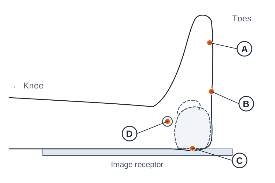
CR: 40° cephalad to the long axis of the foot
Position
Supine or seated, leg extended. Foot dorsiflexed so the plantar surface is near perpendicular to the IR (a strap around the forefoot helps).
Central ray
40° cephalad to the long axis of the foot
Centering
Base of the 3rd metatarsal, so the CR exits through the heel
Typical technique
65–75 kVp · 5–8 mAs · 40" SID
Best shows
Calcaneal body, tuberosity and sustentaculum tali; fractures widening the heel
Image criteria
Entire calcaneus from the tuberosity to the talocalcaneal joint seen.
Calcaneus not foreshortened (too little angle) or elongated (too much).
Sustentaculum tali in profile on the medial side; no rotation.
Lateral calcaneus (mediolateral)
CR: Perpendicular
Position
Lateral recumbent on the affected side, foot dorsiflexed so the plantar surface is perpendicular to the IR.
Central ray
Perpendicular
Centering
About 1 inch (2.5 cm) distal to the medial malleolus
Typical technique
60–65 kVp · 3–5 mAs · 40" SID
Best shows
Calcaneus in profile, the subtalar joint and Böhler's angle (flattened in calcaneal fractures)
Image criteria
Calcaneus seen in lateral profile with the ankle joint and the talus above it.
Sinus tarsi and the cuboid–calcaneal joint open.
No rotation: lateral malleolus over the posterior tibia.
Technique values are typical starting points; follow your department's technique chart.
Labeled diagrams
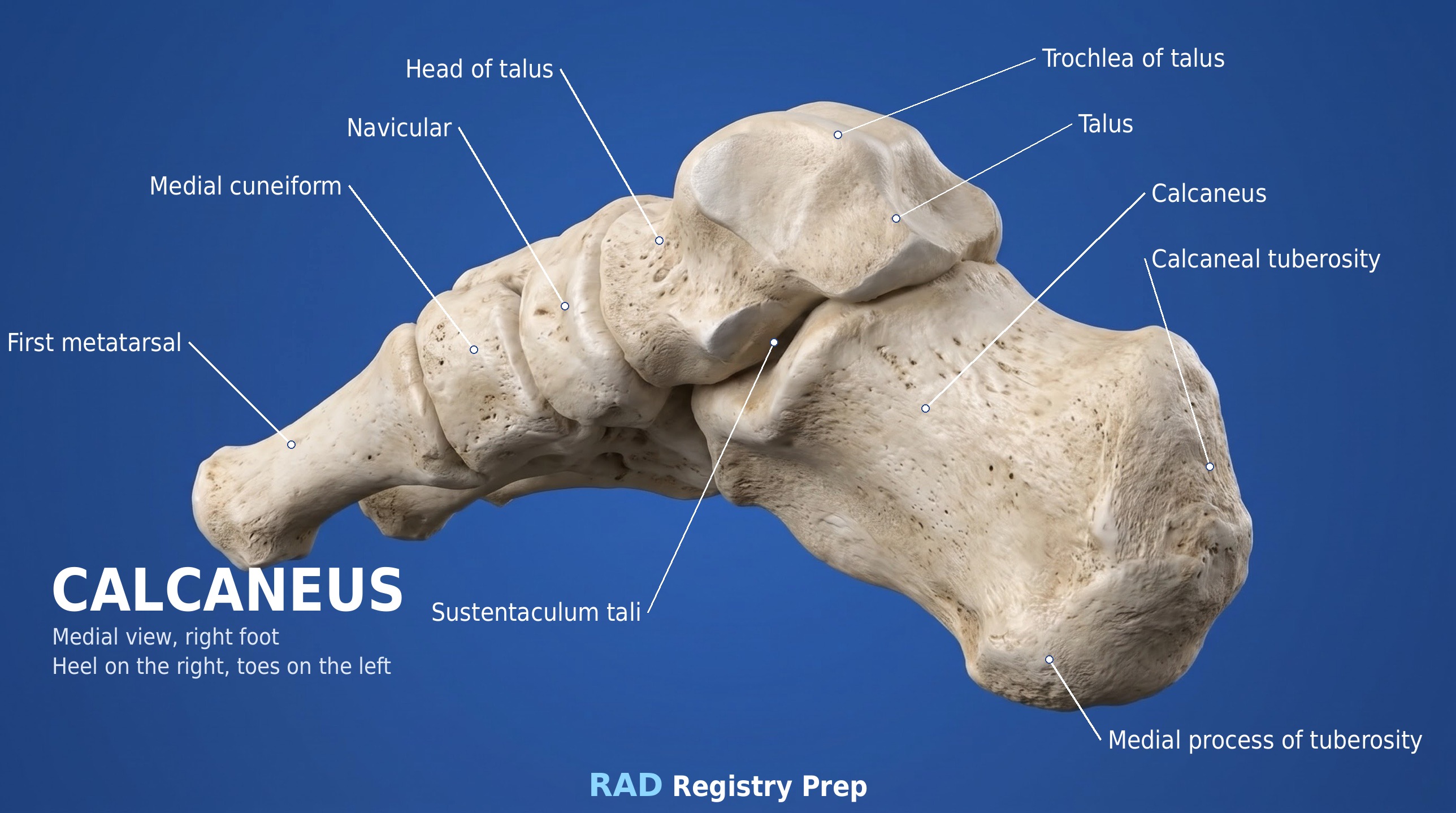
Foot (dorsal view) · Diagram: Rad Registry Prep (original illustration).Calcaneus (medial, right foot (heel on the right) view) · Diagram: Rad Registry Prep (original illustration).
Quick check
1. How is the CR directed for this AP (dorsoplantar) foot?
Image: Mikael Häggström, M.D. CC0 1.0.
2. For bilateral weight-bearing lateral feet to evaluate the longitudinal arches, how is the CR directed?
3. The side-view diagram shows the setup for a plantodorsal axial calcaneus: the patient is supine with the foot dorsiflexed so its long axis is vertical. The CR will be angled 40 degrees cephalad to the long axis of the foot. At which labeled point should the CR enter?
Original diagram: Rad Registry Prep.
4. An AP foot is taken on a 24 x 30 cm field at 100 cm SID. To use the anode heel effect, which part of the foot should be placed toward the cathode end of the tube?
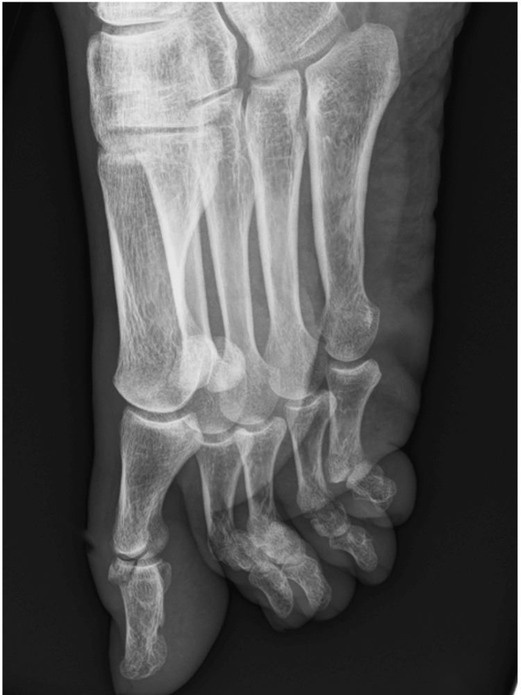
5. This lateral view of the great toe shows a small bone fragment pulled off the dorsal base of the distal phalanx. How is a lateral great toe positioned?
Image: Reis M, Maçães F, Alves B, Gomes M, Macedo S, Cureus 2026, via PubMed Central. CC BY 4.0.