The routine abdomen is a supine AP (KUB). An acute abdomen series adds an erect abdomen (or left lateral decubitus) and often a PA chest to look for free air and air–fluid levels.
Overview
Bones
Bony landmarks
Iliac crest (L4–L5)
Lower ribs
Lumbar spine
Pubic symphysis
ASIS
Soft-tissue structures
Kidneys
Psoas margins
Liver edge
Bladder
Bowel gas pattern
KUB: center at the iliac crest and include the pubic symphysis. Erect abdomen: center about 2 inches above the crest to include the diaphragm.
Registry must-knows
Expose on expiration: the diaphragm rises and the abdomen is less compressed.
Free intraperitoneal air rises under the diaphragm on an erect film; left lateral decubitus shows it over the liver.
Keep the patient erect or on the side for about 5 minutes (10–20 if possible) before an erect or decubitus image.
Routine gonadal shielding is no longer recommended (AAPM 2019, NCRP 2021); follow department policy, and never let a shield cover anatomy of interest.
Show 1 more
Hypersthenic patients may need two IRs placed crosswise.
3D model
Loading 3D…
Radiographs 3
Pick a film, then a structure to circle it.
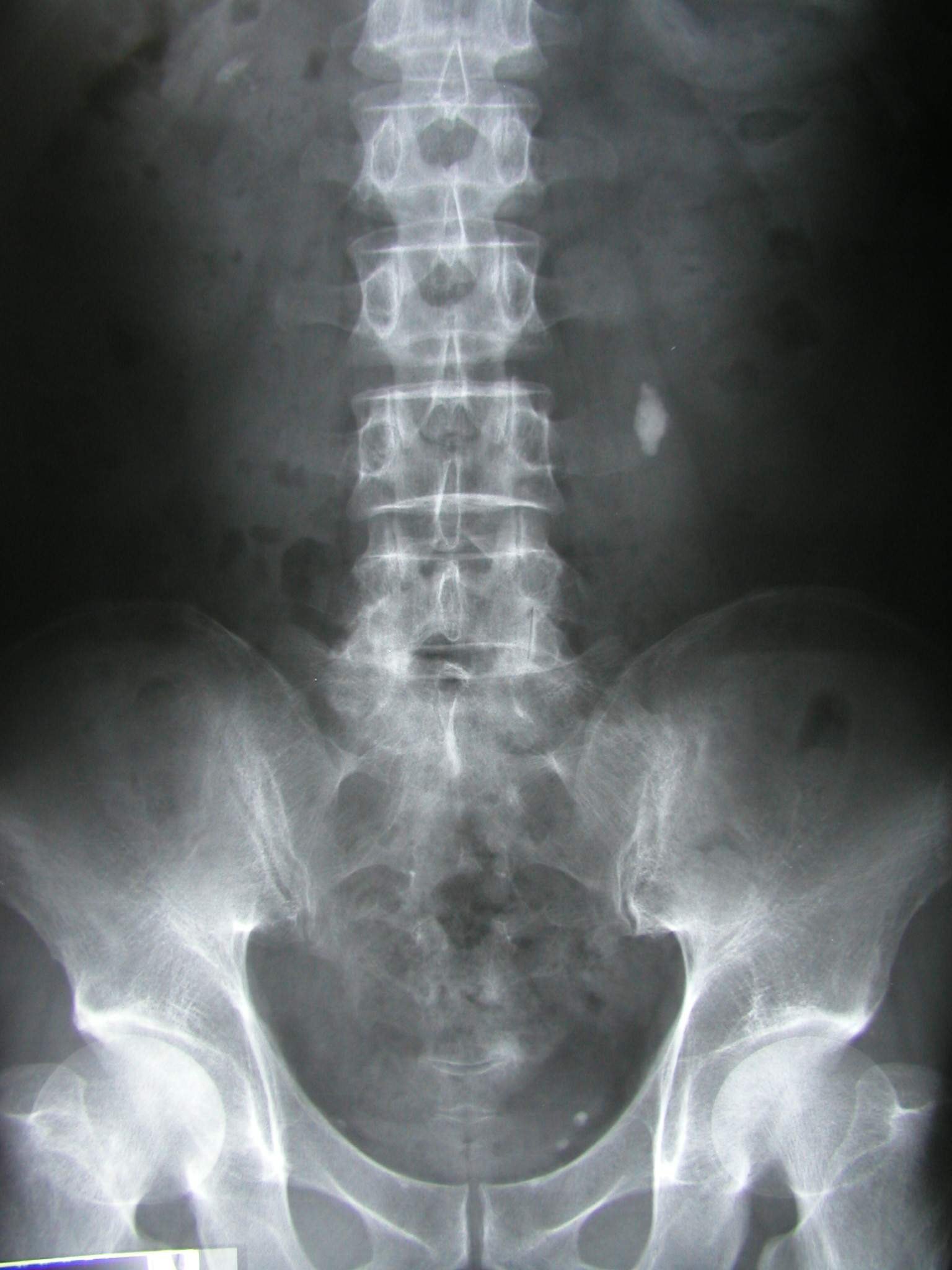
AP abdomen (KUB)Image: Morning2k. CC BY 2.5. Source
Supine, MSP centered, arms at the sides; expose on expiration.
Central ray
Perpendicular
Centering
Iliac crest
Typical technique
75–85 kVp · 20–40 mAs · 40" SID
Image criteria
Pubic symphysis to the upper kidneys included.
No rotation: iliac wings and obturator foramina symmetric.
Psoas margins, kidney outlines and lower liver edge seen.
AP abdomen, erect
CR: Horizontal
Position
Erect, back to the IR, after 5 minutes upright if possible; expiration.
Central ray
Horizontal
Centering
About 2 inches above the iliac crest (to include the diaphragm)
Typical technique
75–85 kVp · 25–50 mAs · 40" SID
Image criteria
Both hemidiaphragms included.
Air–fluid levels shown.
No rotation.
AP abdomen, left lateral decubitus
CR: Horizontal
Position
Lying on the left side for 5 minutes or more; arms up.
Central ray
Horizontal
Centering
About 2 inches above the iliac crest
Typical technique
75–85 kVp · 25–50 mAs · 40" SID
Best shows
Free air over the liver (right side up)
Image criteria
Right side of the abdomen and diaphragm included.
Horizontal beam for air–fluid levels.
Technique values are typical starting points; follow your department's technique chart.
Labeled diagrams
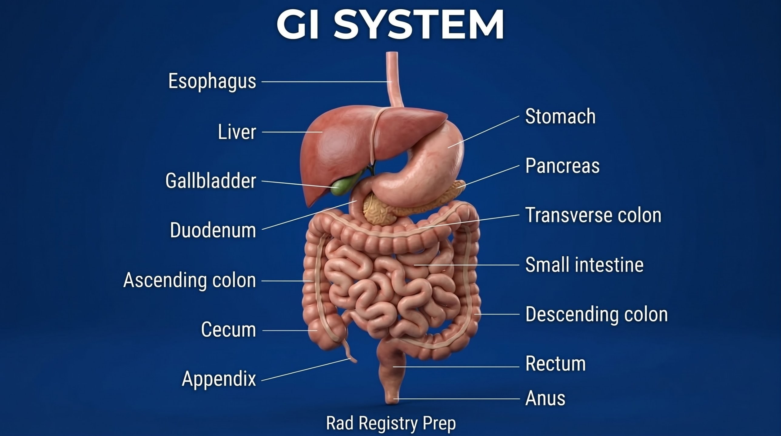
GI System (anterior view) · Diagram: Rad Registry Prep (original illustration).
Quick check
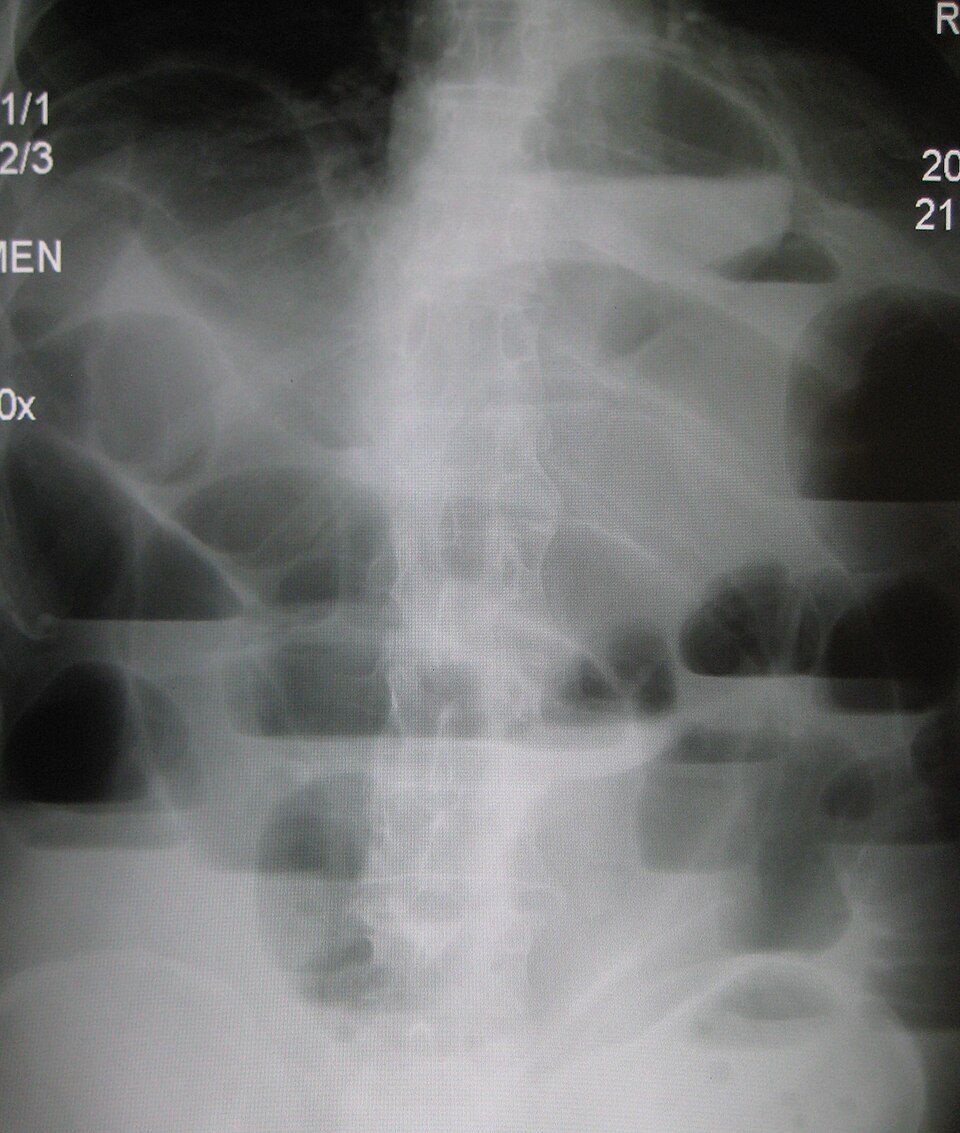
1. The multiple air-fluid levels at different heights in dilated bowel loops on this upright abdomen are MOST consistent with which condition?
Image: James Heilman, MD. CC BY-SA 3.0.
2. This AP supine abdomen on a tall patient includes the pubic symphysis, but the upper poles of the kidneys are above the top edge of the image. What should the radiographer do?
Image: Morning2k. CC BY 2.5.
3. A patient who cannot stand is sent for an acute abdomen series to rule out free air. Which position should replace the upright AP abdomen?
4. An outpatient's upright abdomen series shows a crescent of free air beneath the right hemidiaphragm. The patient says he feels fine and wants to leave. What should the radiographer do?
5. Three days after abdominal surgery, a patient's KUB shows gas-distended loops of both small and large bowel, with gas reaching the rectum and no point of abrupt narrowing. Which condition is MOST likely?