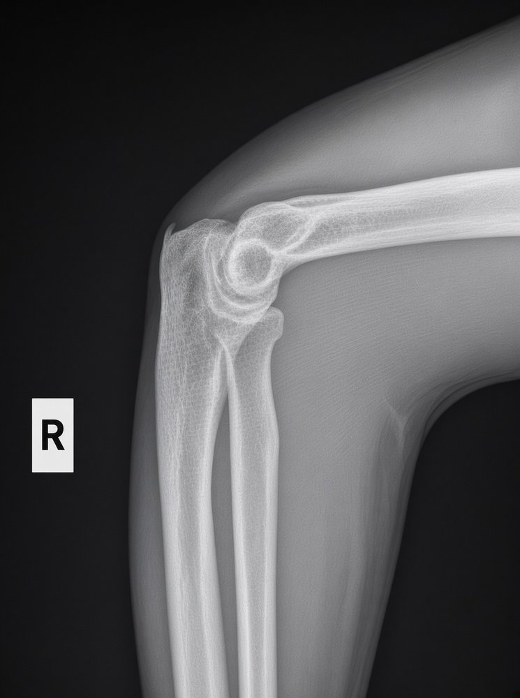
The elbow joins the distal humerus to the radius and ulna. Routine elbow imaging is an AP, a lateral and two obliques; the Coyle methods add axial views of the radial head and coronoid process.
Overview
Bones
Distal humerus
Capitulum (lateral)
Trochlea (medial)
Lateral epicondyle
Medial epicondyle
Olecranon fossa
Coronoid fossa
Proximal radius
Radial head
Radial neck
Radial tuberosity
Proximal ulna
Olecranon
Trochlear notch
Coronoid process
Radial notch
CRITOE, the order the pediatric ossification centers appear: Capitulum, Radial head, Internal (medial) epicondyle, Trochlea, Olecranon, External (lateral) epicondyle.
Registry must-knows
On a lateral with the elbow at 90°, a visible posterior fat pad suggests a joint effusion and an occult fracture.
Radial head fractures are the most common elbow fracture in adults; supracondylar fractures are the most common in children.
External (lateral) oblique frees the radial head, neck and tuberosity from the ulna. Internal (medial) oblique shows the coronoid process in profile.
If the elbow can't extend, take two APs: one with the humerus parallel to the IR and one with the forearm parallel.
Show 1 more
Coyle: elbow 90°, CR 45° toward the shoulder for the radial head; elbow 80°, CR 45° away from the shoulder for the coronoid.
3D model
Loading 3D…
Radiographs 4
Pick a film, then a structure to circle it.
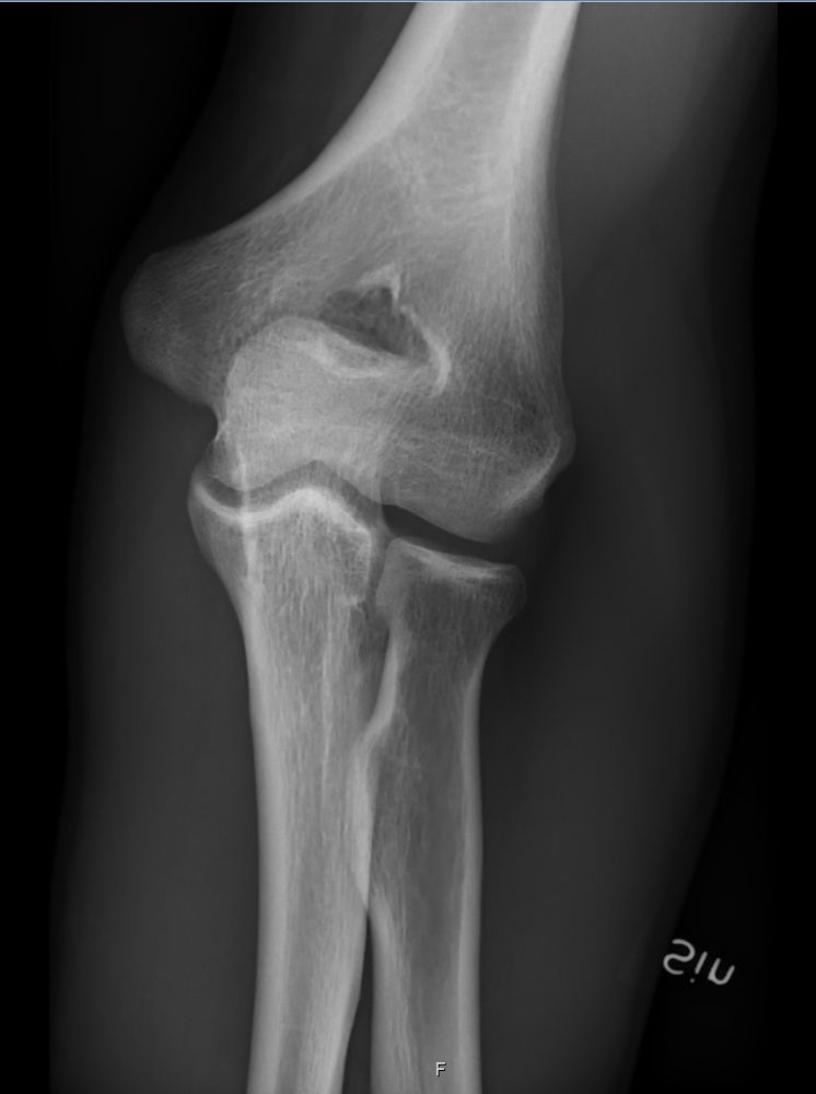
AP elbowImage: Mikael Häggström, M.D., via Wikimedia Commons. CC0 (public domain dedication). Source
Three concentric arcs: trochlear sulcus, double ridges of the capitulum and trochlea, and the trochlear notch.
Radial head partly superimposes the coronoid process; olecranon in profile.
AP oblique, lateral (external) rotation
CR: Perpendicular
Position
From the AP, rotate the hand and arm 45° laterally (hand supinated).
Central ray
Perpendicular
Centering
Mid-elbow joint
Typical technique
60–65 kVp · 3–5 mAs · 40" SID
Best shows
Radial head, neck and tuberosity free of the ulna; capitulum and lateral epicondyle
Image criteria
Radial head, neck and tuberosity free of superimposition.
Capitulum and lateral epicondyle in profile.
Elbow fully extended.
AP oblique, medial (internal) rotation
CR: Perpendicular
Position
From the AP, rotate the hand and arm 45° medially (hand pronated).
Central ray
Perpendicular
Centering
Mid-elbow joint
Typical technique
60–65 kVp · 3–5 mAs · 40" SID
Best shows
Coronoid process in profile; trochlea and medial epicondyle
Image criteria
Coronoid process in profile.
Radial head and neck superimposed by the proximal ulna.
Elbow fully extended.
Axiolateral for the radial head
CoyleCR: 45° toward the shoulder
Position
Elbow flexed 90°, hand pronated.
Central ray
45° toward the shoulder
Centering
Radial head (mid-elbow joint)
Typical technique
65–70 kVp · 4–6 mAs · 40" SID
Best shows
Radial head and capitulum, projected away from the coronoid
Image criteria
Radial head and capitulum free of superimposition.
Joint space between them open.
Axiolateral for the coronoid
CoyleCR: 45° away from the shoulder
Position
Elbow flexed 80°, hand pronated.
Central ray
45° away from the shoulder
Centering
Coronoid process (mid-elbow joint)
Typical technique
65–70 kVp · 4–6 mAs · 40" SID
Best shows
Coronoid process in profile
Image criteria
Coronoid process elongated and in profile.
Distal humerus and coronoid separated.
Technique values are typical starting points; follow your department's technique chart.
Labeled diagrams
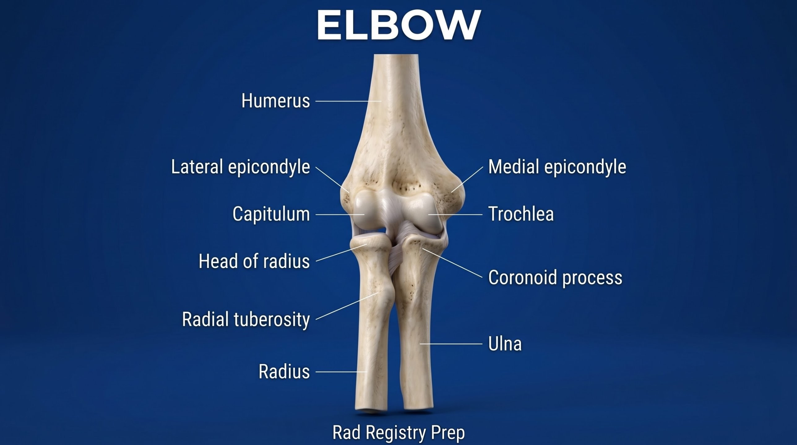
Elbow (anterior view) · Diagram: Rad Registry Prep (original illustration).
Quick check
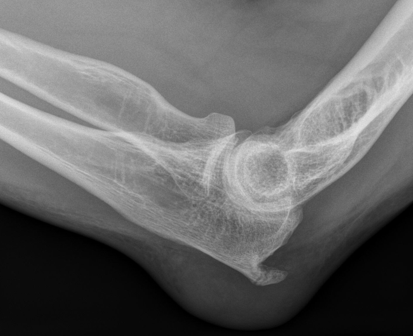
1. The well-defined soft-tissue bulge directly posterior to the olecranon on this lateral elbow radiograph is most consistent with which pathology?
Image: Hellerhoff. CC BY-SA 4.0.
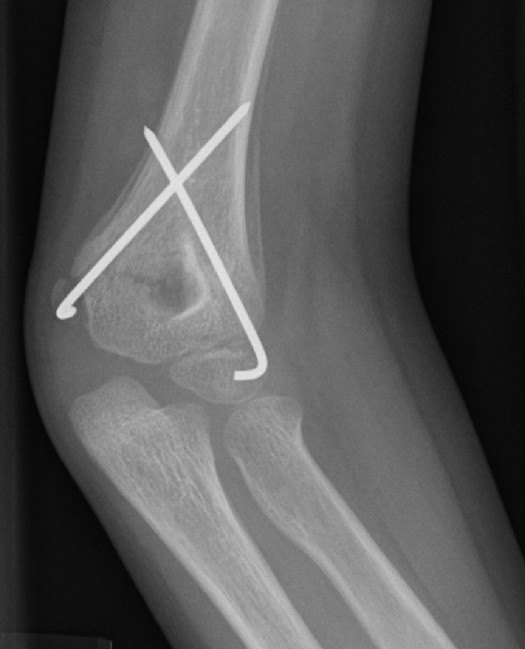
2. This AP elbow of a 6-year-old was obtained after surgery. Two crossed metal pins pass through the distal humerus. Which injury are the pins MOST likely stabilizing?
Image: Lindsaydavidson. CC BY-SA 3.0.
3. A patient cannot fully extend the elbow, which stays flexed about 45°. Two AP projections are needed. For the image of the proximal radius and ulna, how is the arm positioned?
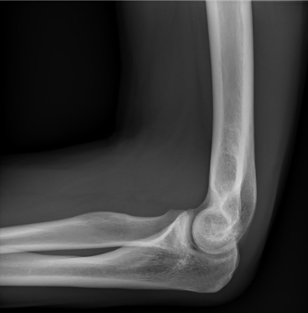
4. How is the elbow positioned for a true lateral elbow like this one?
Image: Langhousen Teaching File, Florida, 2023. Used with permission.
5. Which evaluation criterion shows that this lateral elbow is correctly positioned?
Image: Mikael Häggström, M.D., via Wikimedia Commons. CC0 (public domain dedication).