The forearm is the radius (thumb side) and ulna (little-finger side). A routine forearm is an AP and a lateral, and both must include the wrist and the elbow.
Overview
Bones
Radius (lateral, thumb side)
Head
Neck
Radial tuberosity
Shaft
Styloid process
Ulna (medial, little-finger side)
Olecranon
Coronoid process
Trochlear notch
Shaft
Head
Styloid process
Joints
Proximal radioulnar
Distal radioulnar (DRUJ)
MUGR: Monteggia is an Ulna fracture (with radial head dislocation); Galeazzi is a Radius fracture (with DRUJ dislocation).
Registry must-knows
Always an AP, never a PA: pronating the hand crosses the radius over the ulna.
Include both joints. If the forearm is too long for one IR, add a second image of the other joint.
Monteggia: proximal ulna fracture with a radial head dislocation. Galeazzi: distal radius fracture with a DRUJ dislocation.
A nightstick fracture is an isolated ulnar shaft fracture from a blow to the forearm.
Show 1 more
In children, look for greenstick (one cortex broken) and torus or buckle (cortex bulges) fractures.
3D model
Loading 3D…
Radiographs 2
Pick a film, then a structure to circle it.
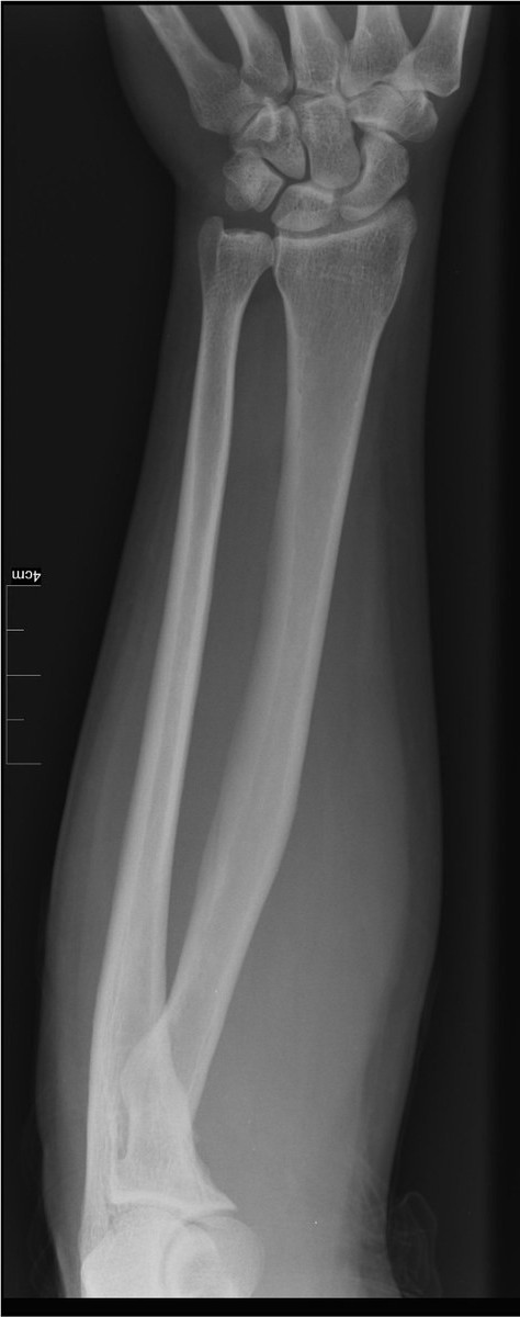
AP forearmImage: Nevit Dilmen, via Wikimedia Commons. CC BY-SA 3.0. Source
Arm extended, hand supinated, epicondyles parallel to the IR. Shoulder lowered to the level of the elbow.
Central ray
Perpendicular
Centering
Mid-forearm
Typical technique
60–65 kVp · 3–4 mAs · 40" SID
Best shows
Radius and ulna side by side, with both joints
Image criteria
Wrist and elbow joints both included.
No rotation: humeral epicondyles in profile; radius and ulna not crossed.
Slight overlap of the radial head, neck and tuberosity on the ulna.
Lateral forearm (lateromedial)
CR: Perpendicular
Position
Elbow flexed 90°, humerus and forearm on the same plane, hand in a true lateral (thumb up).
Central ray
Perpendicular
Centering
Mid-forearm
Typical technique
60–65 kVp · 3–4 mAs · 40" SID
Best shows
Anterior or posterior displacement of fractures
Image criteria
Wrist and elbow joints both included; elbow at 90°.
Distal radius and ulna superimposed; radial head superimposes the coronoid process.
Humeral epicondyles superimposed.
Technique values are typical starting points; follow your department's technique chart.
Labeled diagrams
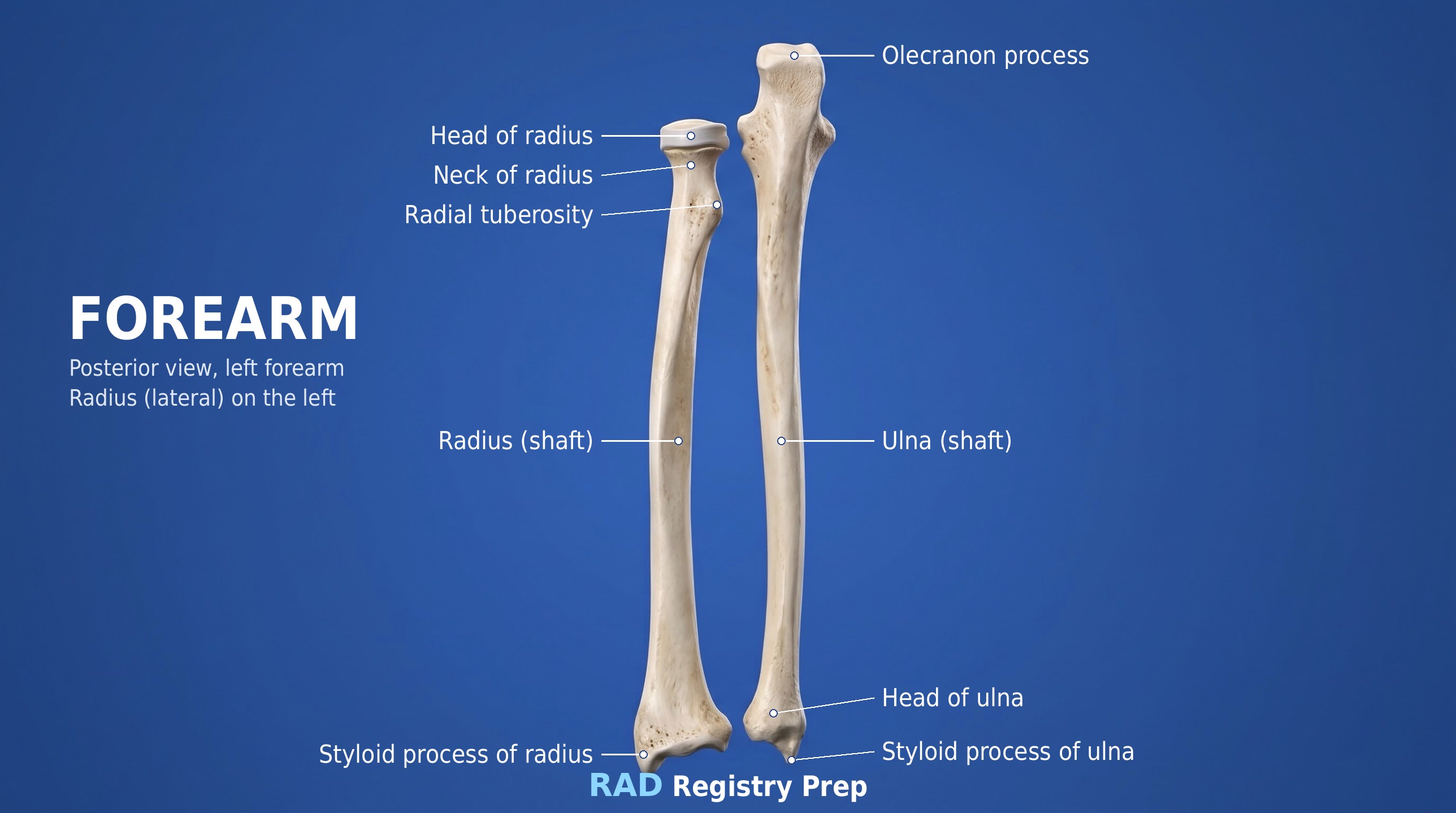
Forearm (posterior, left forearm (radius on the left) view) · Diagram: Rad Registry Prep (original illustration).
Quick check
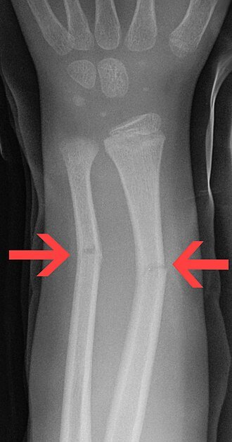
1. A young child has the radius and ulna fractures shown on this forearm radiograph. For the routine AP and lateral forearm projections, which joints must be included?
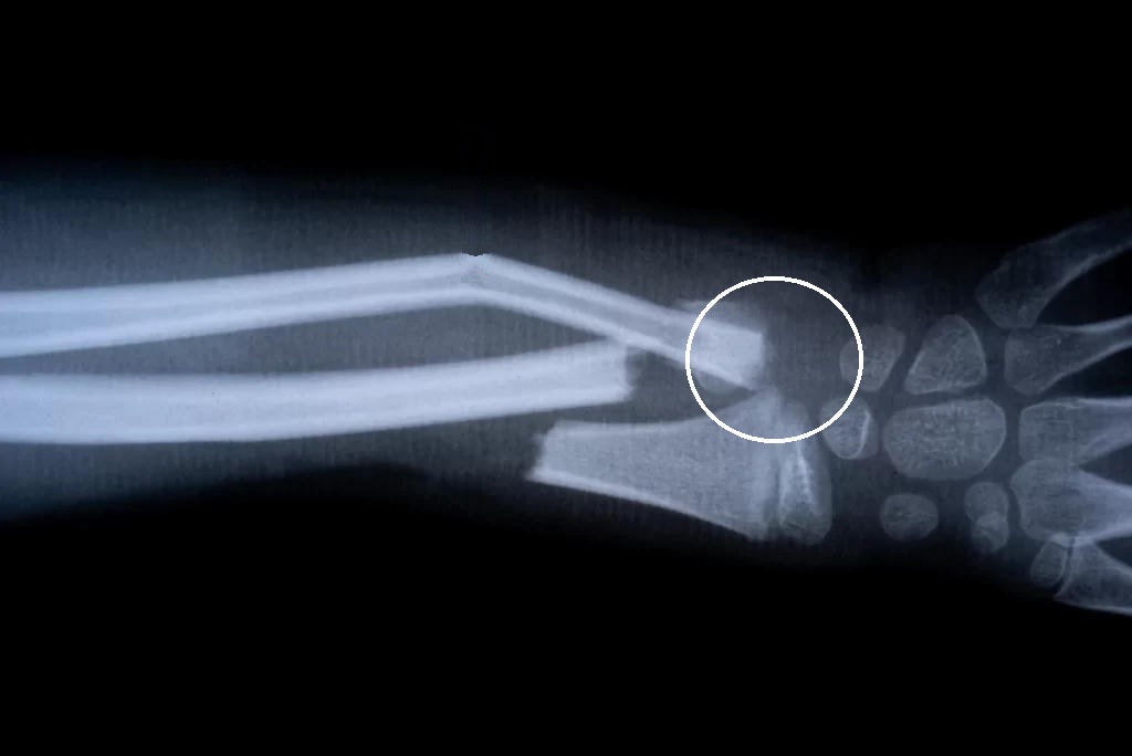
Image: Hellerhoff. CC BY-SA 3.0.
2. Which structure is enclosed by the white circle on this pediatric forearm radiograph?
3. A forearm radiograph shows a fracture of the distal third of the radial shaft. Which associated injury must be checked by including the wrist on both the AP and lateral projections?
4. For this AP forearm, how is the hand positioned?
Image: Nevit Dilmen, via Wikimedia Commons. CC BY-SA 3.0.
5. On an AP forearm, the proximal radius crosses over the ulna. What positioning error caused this, and how should it be corrected?