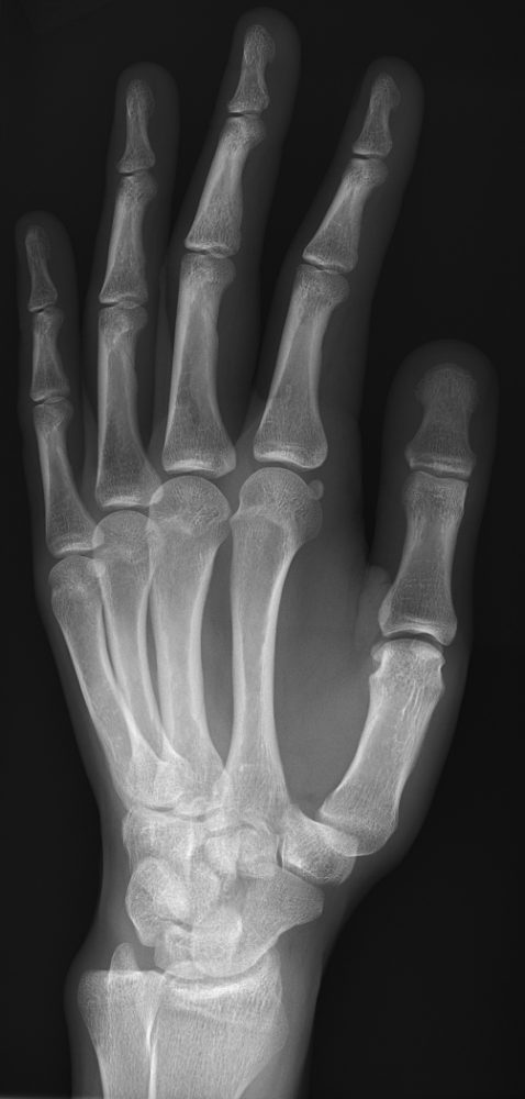
The hand has 27 bones: 14 phalanges, 5 metacarpals and 8 carpals. A routine hand is a PA, a PA oblique and a lateral, centered to the 3rd MCP joint. Fingers and the thumb have their own series (see Fingers & thumb).
Overview
Bones
Phalanges (14)
Thumb: proximal and distal
Fingers 2–5: proximal, middle and distal
Metacarpals (5)
1st (thumb)
2nd
3rd
4th
5th
Joints
IP / PIP / DIP
MCP
CMC
Digits and metacarpals are numbered 1 to 5 starting at the thumb. Each metacarpal has a base (proximal), shaft and head (distal).
Registry must-knows
Centering: hand at the 3rd MCP joint, a finger at its PIP joint, the thumb at the 1st MCP joint.
On the oblique, keep the digits parallel to the IR so the IP joints stay open and the phalanges aren't foreshortened.
Boxer's fracture: the neck of the 5th metacarpal. Bennett fracture: an intra-articular fracture-dislocation at the base of the 1st metacarpal.
Gamekeeper's (skier's) thumb is an ulnar collateral ligament injury at the 1st MCP joint.
Show 1 more
Bone age: a PA of the left hand and wrist. The Norgaard (ball-catcher's) view, both hands 45° AP oblique, looks for early rheumatoid arthritis.
3D model
Loading 3D…
Radiographs 4
Pick a film, then a structure to circle it.
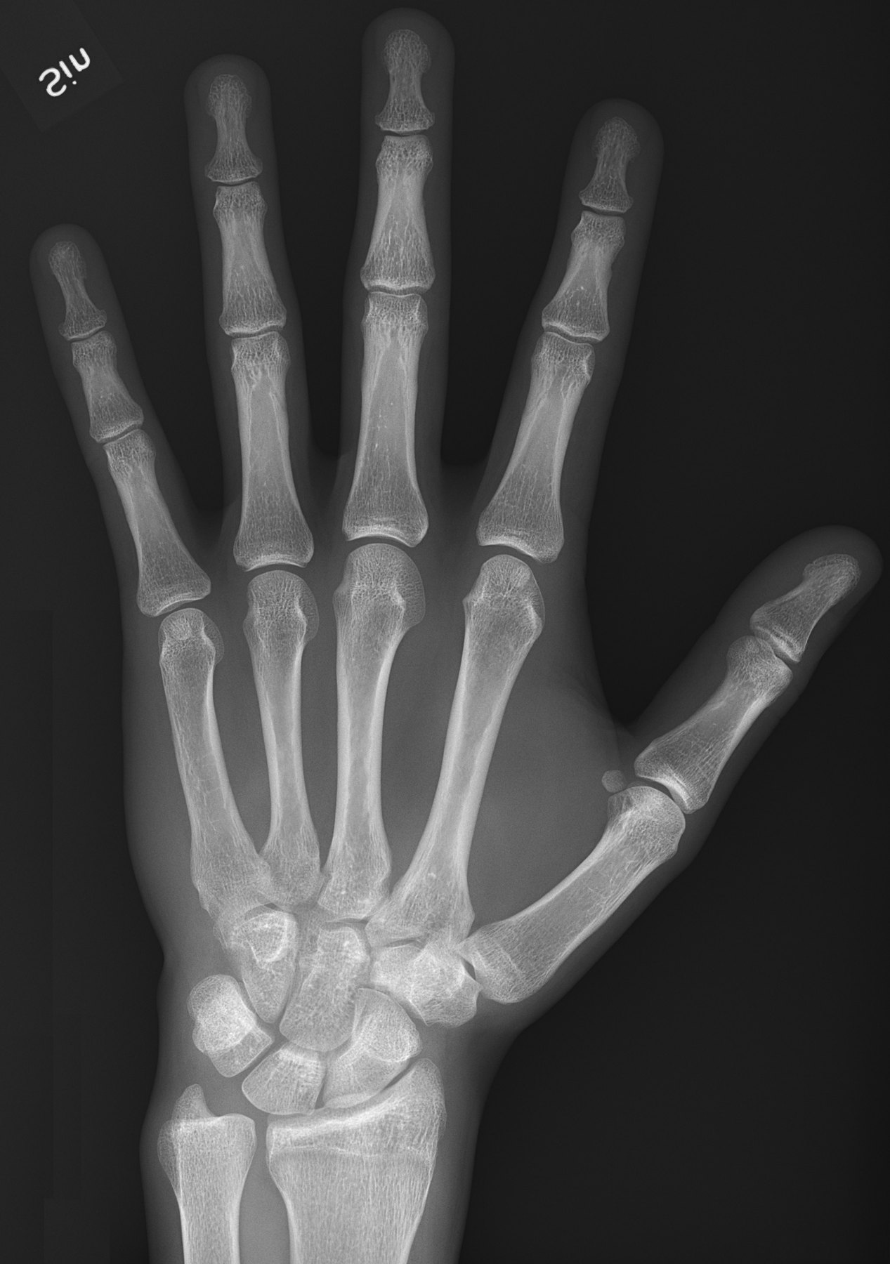
PA handImage: Mikael Häggström, M.D., via Wikimedia Commons. CC0 (public domain dedication). Source
All the bones of the hand; the thumb appears oblique
Image criteria
Entire hand and about 1 inch of the distal radius and ulna included.
No rotation: equal concavity on both sides of the metacarpal and phalangeal shafts.
Digits slightly separated; MCP and IP joints open.
PA oblique hand (lateral rotation)
CR: Perpendicular
Position
From the PA, rotate the hand 45° laterally, with the digits supported parallel to the IR.
Central ray
Perpendicular
Centering
3rd MCP joint
Typical technique
55–60 kVp · 2–3 mAs · 40" SID
Best shows
Metacarpal and phalangeal fractures; joint spaces
Image criteria
Midshafts of the 3rd–5th metacarpals don't overlap; slight overlap of their heads is normal.
IP and MCP joints open (digits parallel to the IR).
No more than 45° of rotation.
Lateral hand
Fan lateralCR: Perpendicular
Position
Ulnar side down, hand in a true lateral. Fan the fingers on a step sponge to see the phalanges, or keep them together (extension) for a foreign body or metacarpal displacement.
Central ray
Perpendicular
Centering
2nd MCP joint
Typical technique
55–60 kVp · 2–4 mAs · 40" SID
Best shows
Anterior or posterior displacement of metacarpal fractures; foreign bodies
Image criteria
Metacarpals superimposed; distal radius and ulna superimposed.
Fan lateral: each phalanx seen separately, IP joints open.
Thumb free of the other digits.
Technique values are typical starting points; follow your department's technique chart.
Labeled diagrams
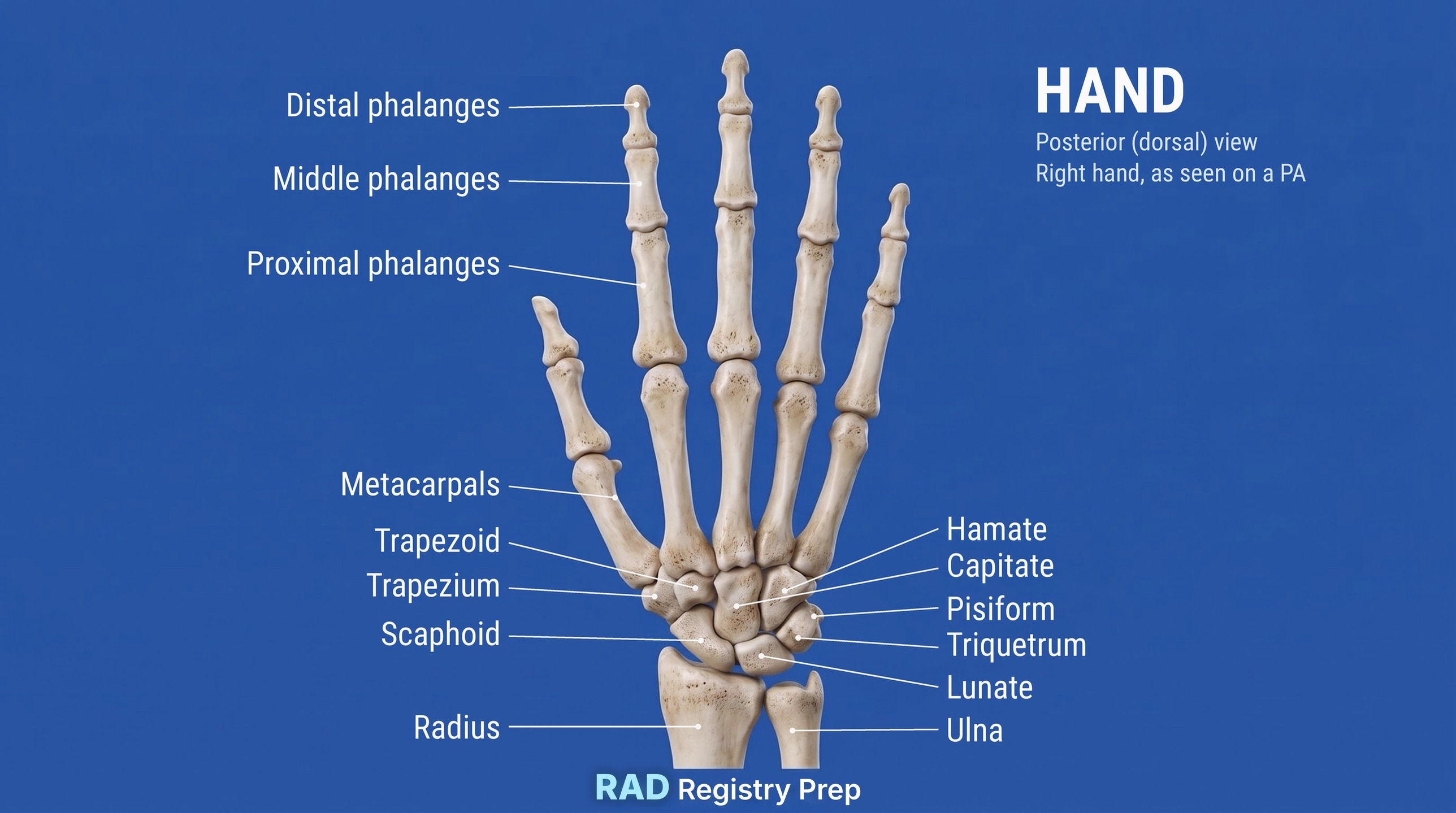
Hand (posterior (dorsal), right hand as seen on a PA view) · Diagram: Rad Registry Prep (original illustration).
Quick check
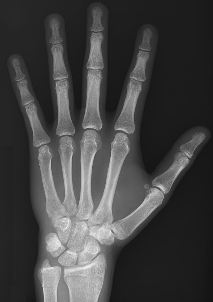
1. On this PA hand, the interphalangeal and metacarpophalangeal joints appear open and the phalanges are not foreshortened. Which positioning step produced this?
Image: Mikael Häggström, M.D. CC0 1.0.
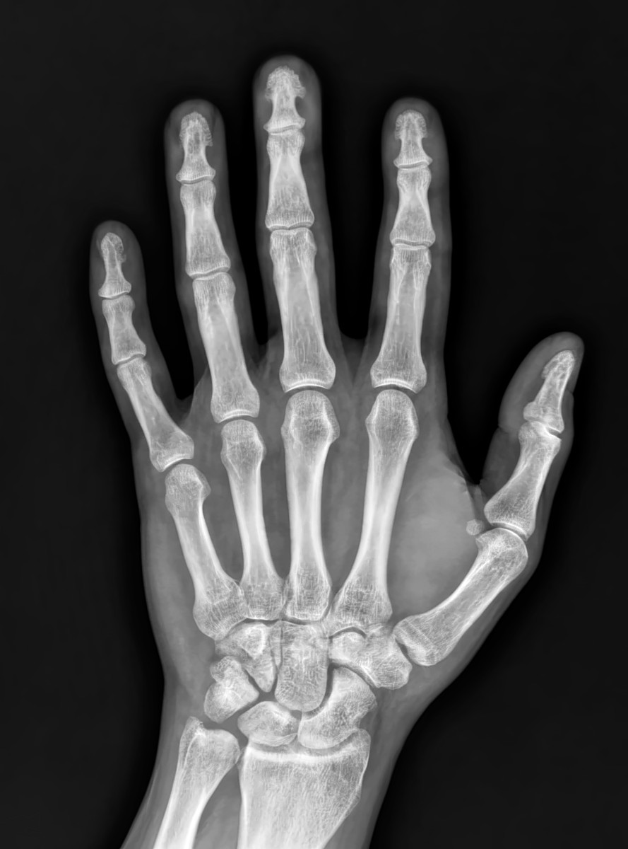
2. A PA hand such as this one is usually performed on the tabletop without a grid. What is the MAIN reason?
Image: Langhousen teaching library, 2023.
3. On a PA oblique hand, the midshafts of the third through fifth metacarpals overlap. What positioning error caused this?
4. A skier fell with the thumb caught in the ski pole strap and now has pain on the ulnar side of the thumb's MCP joint. Gamekeeper's (skier's) thumb is an injury of which structure?
5. On a PA oblique hand, the interphalangeal joints of the fingers appear closed and the phalanges look foreshortened. What is the MOST likely cause?
Image: Mikael Häggström, M.D., via Wikimedia Commons. CC0 (public domain dedication).