The humerus runs from the shoulder to the elbow. A routine humerus is an AP and a rotational lateral that include both joints; trauma patients get a transthoracic lateral instead of rotating the arm.
Overview
Bones
Proximal
Head
Anatomical neck
Surgical neck
Greater tubercle
Lesser tubercle
Intertubercular (bicipital) groove
Shaft
Deltoid tuberosity
Radial groove
Distal
Capitulum
Trochlea
Medial epicondyle
Lateral epicondyle
Olecranon fossa
Coronoid fossa
Registry must-knows
If a fracture is suspected, don't rotate the arm. Use a transthoracic lateral (Lawrence) or a scapular Y.
AP: epicondyles parallel to the IR puts the greater tubercle in profile laterally. Lateral: epicondyles perpendicular puts the lesser tubercle in profile medially.
The surgical neck is a common fracture site in older adults after a fall.
The radial nerve runs in the radial groove, so it can be injured with a midshaft fracture.
Show 1 more
Include both the shoulder and elbow joints; use a second image if the IR is too short.
3D model
Loading 3D…
Radiographs 3
Pick a film, then a structure to circle it.
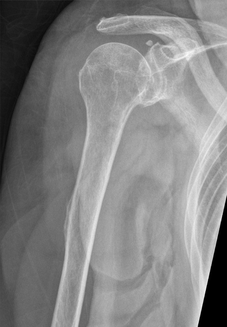
AP humerusImage: Hellerhoff, via Wikimedia Commons. CC BY-SA 4.0. Source
Erect or supine, arm slightly abducted, hand supinated, epicondyles parallel to the IR.
Central ray
Perpendicular
Centering
Mid-humerus
Typical technique
70–75 kVp · 6–10 mAs · 40" SID
Image criteria
Shoulder and elbow joints included.
Epicondyles in profile; greater tubercle in profile laterally.
Humeral head partly superimposes the glenoid.
Lateral humerus (rotational)
CR: Perpendicular
Position
Rotate the arm internally (back of the hand against the hip), epicondyles perpendicular to the IR.
Central ray
Perpendicular
Centering
Mid-humerus
Typical technique
70–75 kVp · 6–10 mAs · 40" SID
Image criteria
Shoulder and elbow joints included.
Epicondyles superimposed; lesser tubercle in profile medially.
Do not use when a fracture is suspected.
Transthoracic lateral
LawrenceCR: Perpendicular (10–15° cephalad if the opposite shoulder can't be raised)
Position
Affected arm against the IR in neutral rotation; opposite arm raised over the head. Breathing technique blurs the ribs and lung markings.
Central ray
Perpendicular (10–15° cephalad if the opposite shoulder can't be raised)
Centering
Surgical neck
Typical technique
75–85 kVp · 20–40 mAs · 40" SID
Best shows
Proximal humerus seen through the thorax, for trauma
Image criteria
Proximal humerus seen through the thorax, clear of the opposite humerus.
Ribs and lung markings blurred by breathing.
Technique values are typical starting points; follow your department's technique chart.
Labeled diagrams
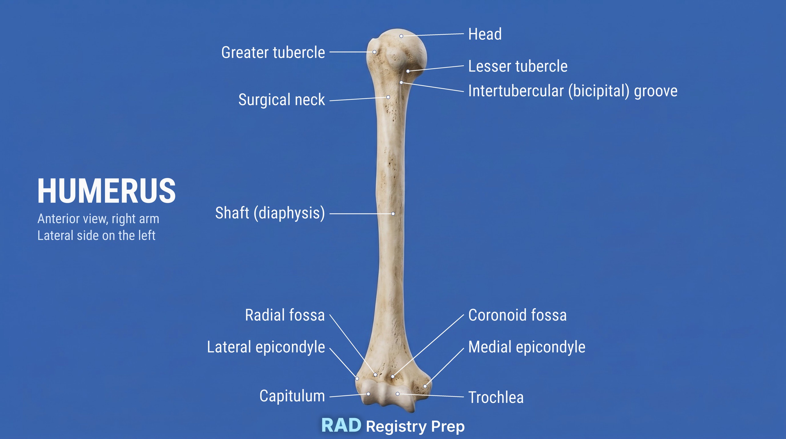
Humerus (anterior, right arm (lateral side on the left) view) · Diagram: Rad Registry Prep (original illustration).
Quick check
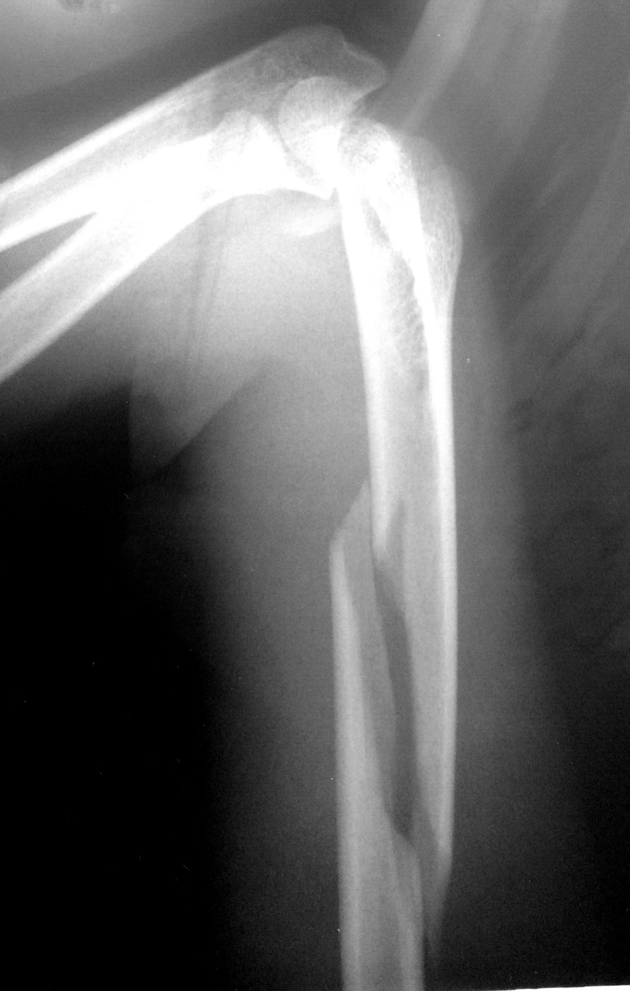
1. This radiograph of the upper arm was taken after a fall. The flexed elbow is at the top of the image. Which description BEST fits the injury?
Image: Ivtorov. CC BY-SA 4.0.
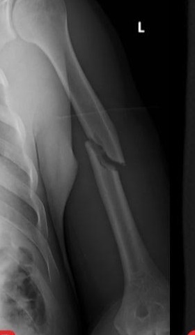
2. This patient's humeral fracture prevents any rotation or abduction of the arm. Which projection should the radiographer use to obtain a lateral view of the proximal humerus?
3. For a non-trauma AP projection of the humerus, how should the humeral epicondyles be positioned?
4. On this AP humerus, the greater tubercle is seen in profile on the lateral side of the humeral head. How was the arm positioned?
Image: Hellerhoff, via Wikimedia Commons. CC BY-SA 4.0.
5. An AP shoulder projection is taken with the hand supinated and the humeral epicondyles parallel to the digital detector. Which structure is placed in profile on the lateral side of the proximal humerus?