Each finger has three phalanges (proximal, middle and distal) and the thumb has two. A finger series is a PA, a PA oblique and a lateral centered to the PIP joint; the thumb series is an AP (or PA), a lateral and an oblique centered to the 1st MCP joint.
Overview
Bones
Fingers 2–5 (3 phalanges each)
Proximal phalanx
Middle phalanx
Distal phalanx (tuft at the tip)
Thumb (2 phalanges)
Proximal phalanx
Distal phalanx
1st metacarpal
Joints
DIP and PIP (fingers)
IP (thumb)
MCP
1st CMC (base of the thumb)
Digits are numbered 1 to 5 from the thumb. Fingers are centered to the PIP joint; the thumb to the 1st MCP joint.
Registry must-knows
Include the distal half of the metacarpal on every finger and thumb view so a fracture at the base isn't missed.
Keep the finger parallel to the IR on the oblique and lateral; if it tilts, the IP joints close and the phalanges foreshorten.
Lateral finger: 2nd digit radial side down (less OID), 3rd–5th ulnar side down; the other fingers are curled into a fist out of the way.
The AP thumb (back of the thumb on the IR) has less OID than the PA thumb, so it is sharper; the PA is used when the patient can't rotate the hand.
Show 1 more
Mallet finger: avulsion of the extensor tendon at the base of the distal phalanx, best seen on the lateral. Bennett and Rolando fractures are at the base of the 1st metacarpal; gamekeeper's thumb is the ulnar collateral ligament of the 1st MCP joint.
3D model
Loading 3D…
Radiographs 5
Pick a film, then a structure to circle it.
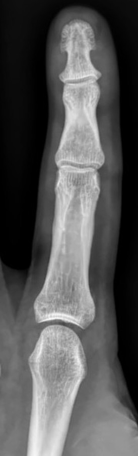
PA finger (2nd digit), cropped from a PA handLabels for this film are coming.Image: Langhousen teaching library, 2023.
Palm flat on the IR, the affected finger extended and slightly separated from the others.
Central ray
Perpendicular
Centering
PIP joint of the affected finger
Typical technique
50–55 kVp · 1–2 mAs · 40" SID
Best shows
The phalanges of one finger and the distal metacarpal, with the IP and MCP joints
Image criteria
Whole digit and the distal half of the metacarpal included.
No rotation: equal concavity on both sides of the phalangeal shafts.
IP and MCP joints open.
PA oblique finger (45° lateral rotation)
CR: Perpendicular
Position
From the PA, rotate the hand 45° laterally (thumb side up) with the finger resting on a 45° sponge, parallel to the IR. The 2nd digit may be rotated medially instead to cut OID.
Central ray
Perpendicular
Centering
PIP joint
Typical technique
50–55 kVp · 1–2 mAs · 40" SID
Best shows
Phalangeal fractures and IP joints not seen on the PA
Image criteria
Digit parallel to the IR: IP joints open, phalanges not foreshortened.
More concavity on one side of the phalangeal shafts.
Distal metacarpal included.
Lateral finger
CR: Perpendicular
Position
True lateral with the affected finger extended and the others flexed into a fist. 2nd digit: radial side to the IR (mediolateral). 3rd–5th digits: ulnar side to the IR (lateromedial). Support the finger on a sponge so it is parallel to the IR.
Central ray
Perpendicular
Centering
PIP joint
Typical technique
50–55 kVp · 1–2 mAs · 40" SID
Best shows
Anterior or posterior displacement of phalangeal fractures; the IP joints in profile
Image criteria
Finger in a true lateral, free of the other digits.
IP joints open.
Concave anterior surfaces of the phalanges; distal metacarpal included.
AP thumb
Roberts (AP axial)CR: Perpendicular to the 1st MCP joint. For Roberts, angle 15° proximally and center to the 1st CMC joint.
Position
Rotate the hand internally until the back of the thumb rests on the IR.
Central ray
Perpendicular to the 1st MCP joint. For Roberts, angle 15° proximally and center to the 1st CMC joint.
Centering
1st MCP joint (1st CMC joint for Roberts)
Typical technique
50–55 kVp · 1–2 mAs · 40" SID
Best shows
The first metacarpal and phalanges; Roberts opens the 1st CMC joint (Bennett fracture, arthritis)
Image criteria
Trapezium through the distal phalanx included.
No rotation: equal concavity of the shafts.
Joints open; soft tissue of other digits not over the thumb.
Lateral thumb
CR: Perpendicular
Position
From the PA hand position, flex the hand slightly so the thumb lies in a true lateral.
Central ray
Perpendicular
Centering
1st MCP joint
Typical technique
50–55 kVp · 1–2 mAs · 40" SID
Image criteria
Thumb in a true lateral, free of the other digits.
Concave anterior surfaces of the proximal phalanx and metacarpal.
IP and MCP joints open.
Technique values are typical starting points; follow your department's technique chart.
Labeled diagrams
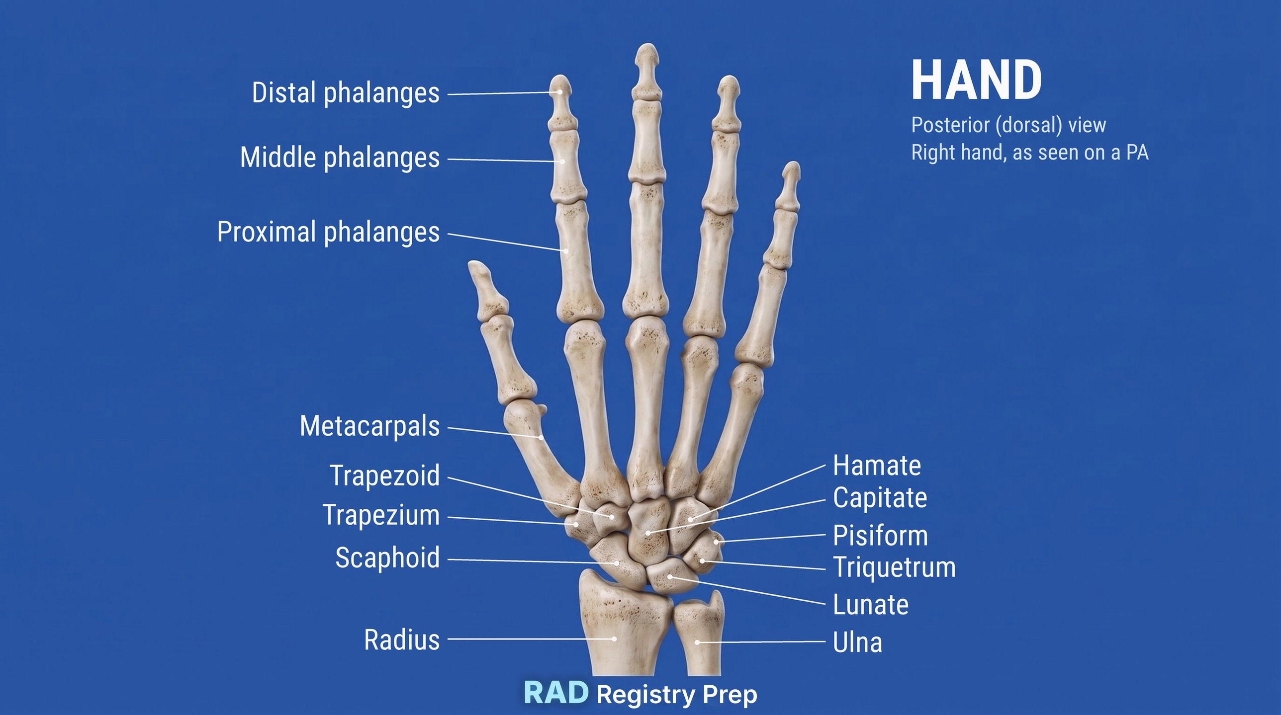
Hand (posterior (dorsal), right hand as seen on a PA view) · Diagram: Rad Registry Prep (original illustration).
Quick check
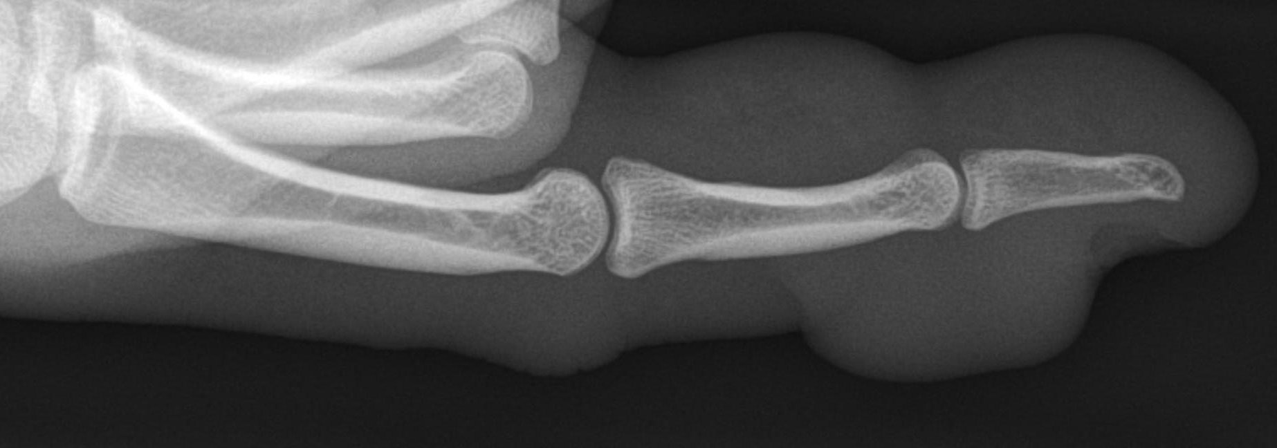
1. The enlarged, lobulated soft-tissue outline surrounding intact bones on this lateral thumb radiograph is most consistent with which finding?
Image: Hellerhoff. CC BY-SA 4.0.
2. A PA projection of the third finger is ordered, and the room's table Bucky is set up for AEC. The finger is much narrower than the AEC chamber. Which approach is MOST appropriate?
3. For an AP projection of the thumb, the hand is internally rotated so the posterior surface of the thumb rests on the digital detector. Where is the CR directed?
4. A patient jammed a fingertip while catching a ball and now cannot straighten the tip of the finger (mallet finger). Which projection BEST shows a small avulsion fragment from the dorsal base of the distal phalanx?
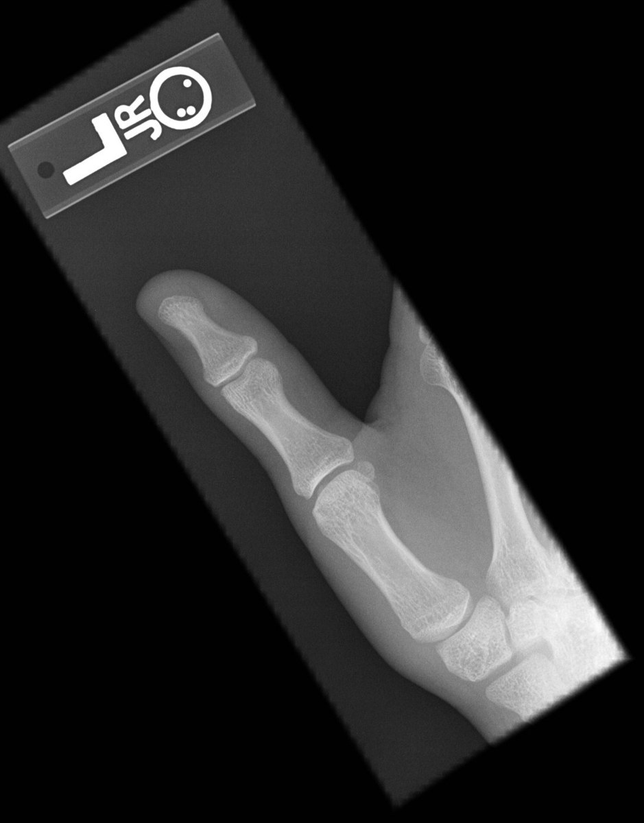
5. This PA thumb was taken with the palm on the IR. Why is the AP thumb usually preferred over the PA?
Image: Staff at the Department of Radiology, UC San Diego Health, via Wikimedia Commons. Public domain.