The lower leg is the tibia (medial, weight-bearing) and fibula (lateral). A routine tib-fib is an AP and a lateral that include both the knee and the ankle.
Overview
Bones
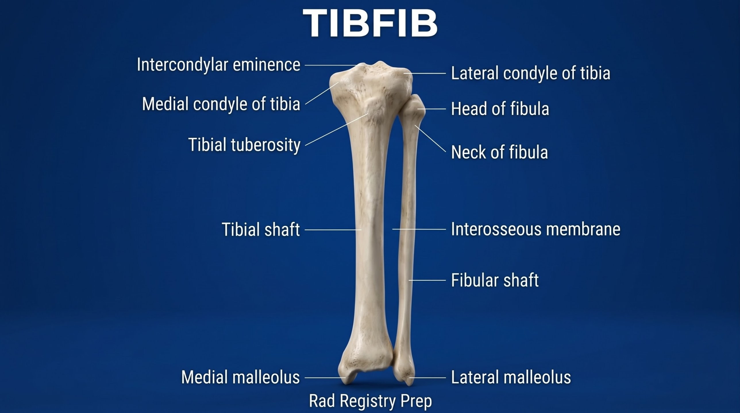
Tibia
Medial condyle
Lateral condyle
Intercondylar eminence
Tibial tuberosity
Shaft (anterior crest)
Medial malleolus
Fibula
Head
Neck
Shaft
Lateral malleolus
Registry must-knows
Include both joints. Place the IR diagonally or use two images if the leg is too long.
Use the anode heel effect: cathode over the thicker knee end.
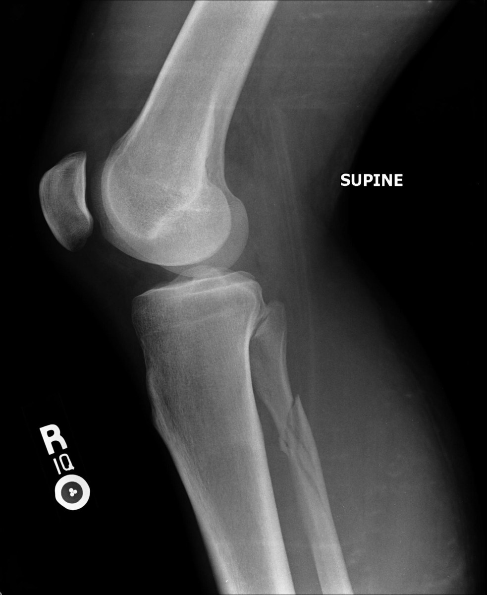
Maisonneuve fracture: a proximal fibula fracture with a medial ankle injury, so image the whole leg after an ankle injury with medial tenderness.
The tibia is the weight-bearing bone; the fibula carries little load.
3D model
Loading 3D…
Radiographs 2
Pick a film, then a structure to circle it.
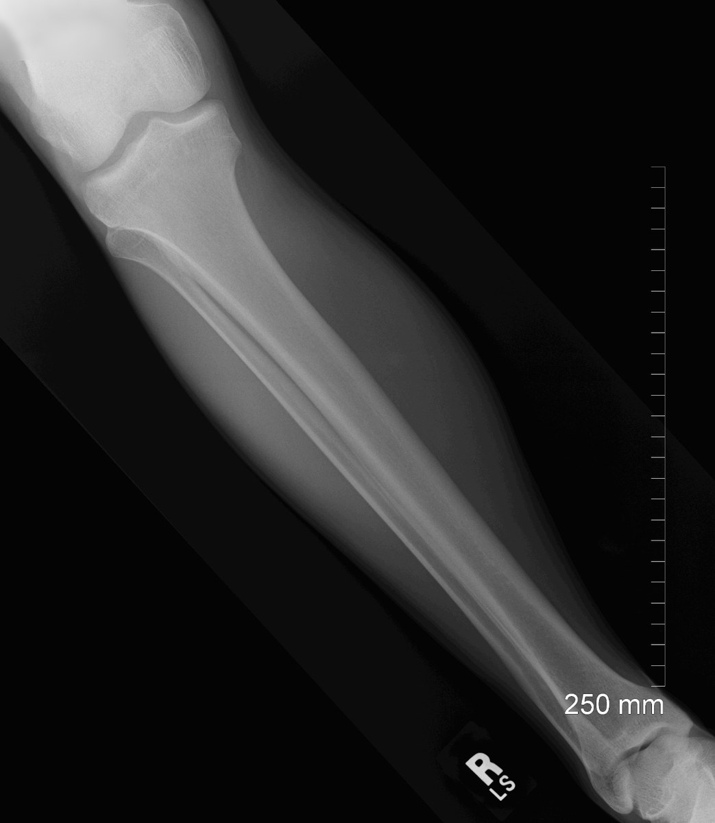
AP tibia & fibulaImage: Eric Schmuttenmaer, via Wikimedia Commons. CC BY-SA 2.0. Source
Supine, leg extended, femoral condyles parallel to the IR, foot dorsiflexed.
Central ray
Perpendicular
Centering
Mid-leg
Typical technique
65–70 kVp · 4–8 mAs · 40–44" SID
Image criteria
Knee and ankle joints both included.
No rotation: femoral and tibial condyles symmetric.
Slight overlap of the tibia and fibula at both ends.
Lateral tibia & fibula (mediolateral)
CR: Perpendicular
Position
Lateral recumbent on the affected side, knee flexed about 45°, femoral condyles superimposed.
Central ray
Perpendicular
Centering
Mid-leg
Typical technique
65–70 kVp · 4–8 mAs · 40–44" SID
Image criteria
Knee and ankle joints both included.
Fibula partly superimposed by the tibia at both ends.
Femoral condyles superimposed.
Technique values are typical starting points; follow your department's technique chart.
Labeled diagrams
Tibia and Fibula (anterior view) · Diagram: Rad Registry Prep (original illustration).
Quick check
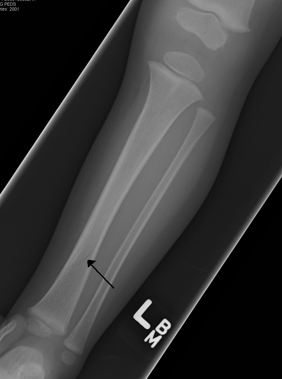
1. This lower-leg image of a toddler shows a subtle fracture of the distal tibial shaft (arrow). The leg was placed diagonally across the detector. What is the MAIN reason for this placement?
Image: James Heilman, MD. CC BY-SA 3.0.
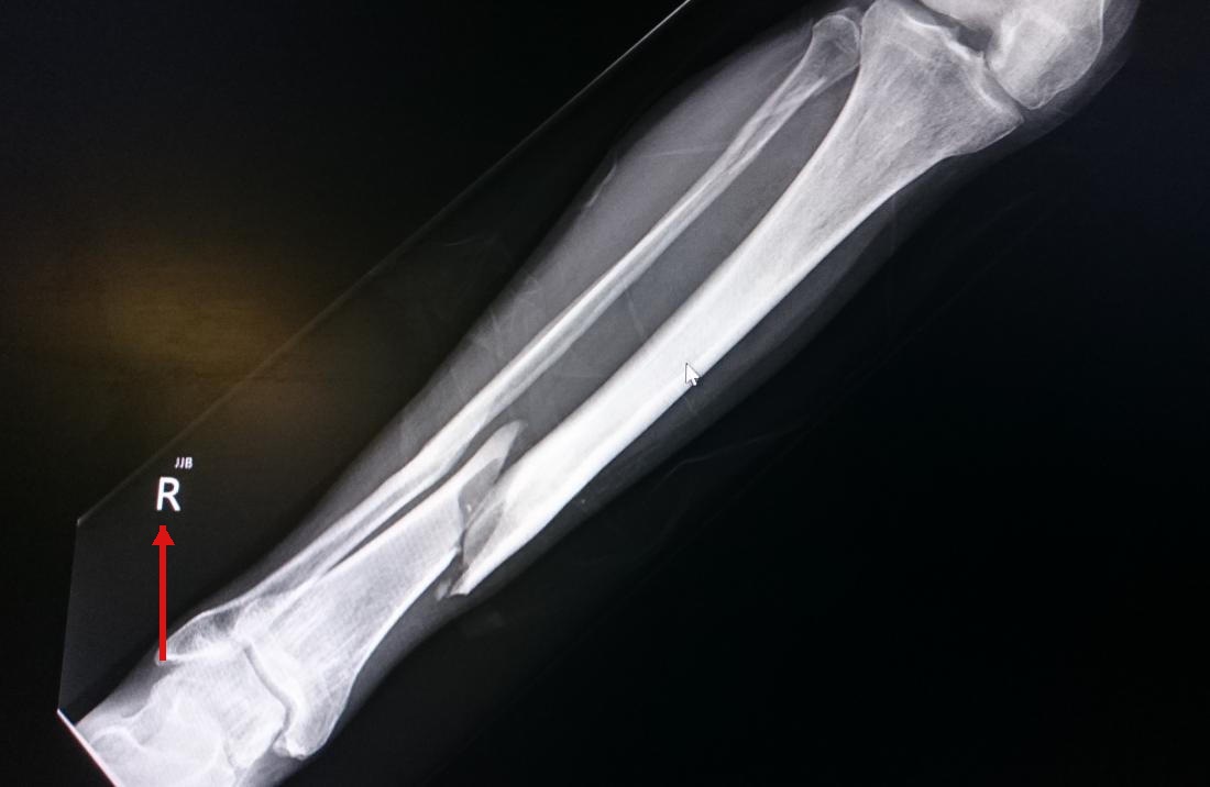
2. The red arrow points to the anatomic side marker on this tibia-fibula radiograph. Where should this marker be placed?
3. A 7-year-old has fever and a painful, swollen lower leg, and acute osteomyelitis of the distal tibia is suspected. Radiographs taken 2 days after symptoms began show no bone abnormality. What is the BEST explanation?
4. This lateral knee shows a fracture of the proximal fibular shaft in a patient who rolled the ankle. This combination, a Maisonneuve fracture, is associated with injury to which structures?
Image: RotorMotor2, via Wikimedia Commons. CC BY-SA 3.0.