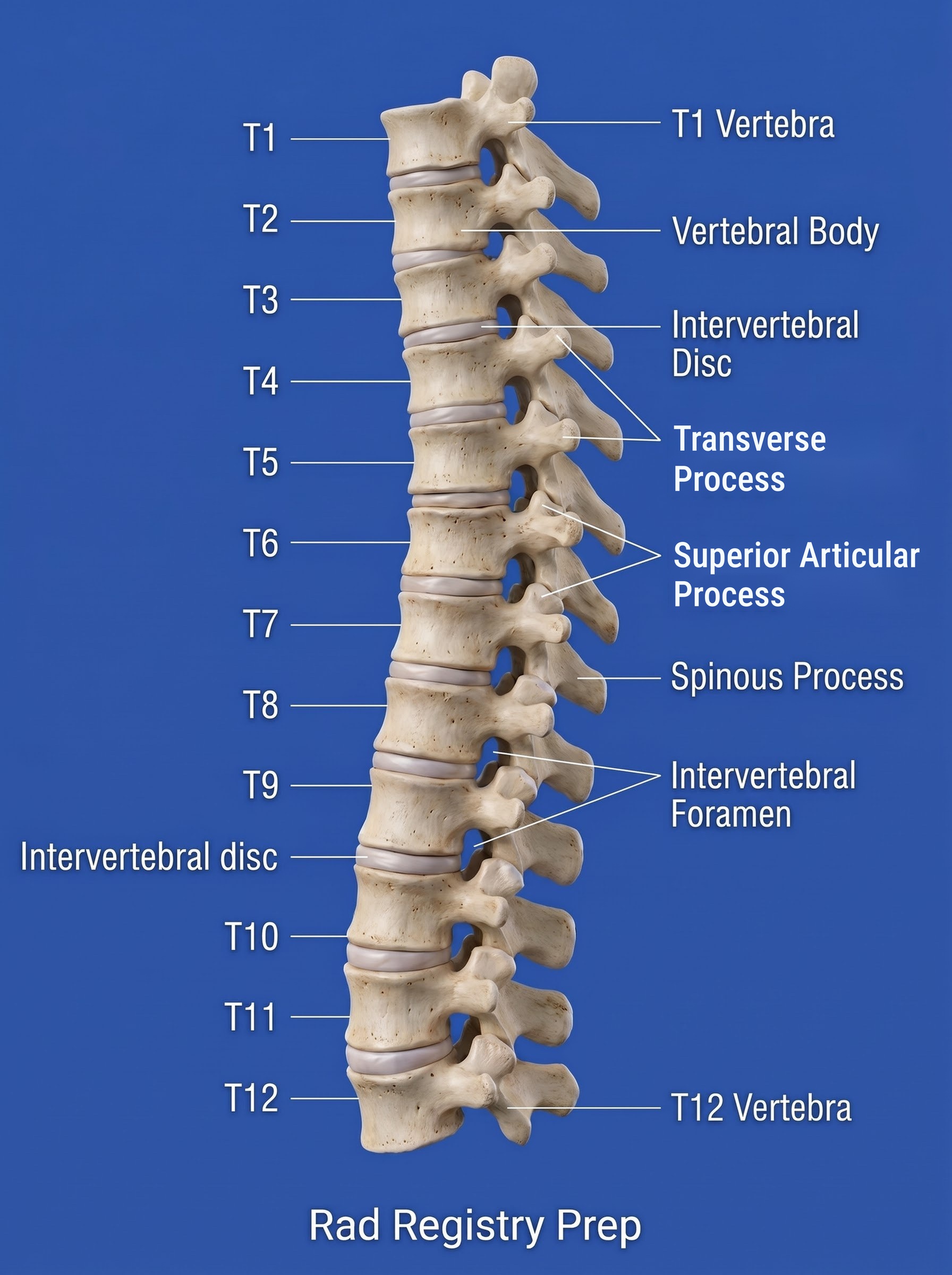
The thoracic spine has 12 vertebrae, each with facets for the ribs. A routine T-spine is an AP and a lateral; the lateral uses a breathing technique to blur the ribs and lungs. The scoliosis series (a standing long-length frontal and lateral of the whole spine) is one of the most common spine exams and is shown on this page too.
Overview
Bones
Thoracic vertebrae
Body with costal facets
Long, downward spinous processes
Transverse processes with costal facets
Zygapophyseal joints (best on 70° obliques)
T7 is about 3–4 inches below the jugular notch (or 1–2 inches below the sternal angle), the center of a T-spine.
Registry must-knows
AP: use the anode heel effect — cathode over the thicker lower T-spine.
Lateral: breathing technique (orthostatic) with a long exposure time blurs the ribs and lung markings.
Zygapophyseal joints of the T-spine open on 70° obliques (20° from lateral).
Collimate tightly and place a lead blocker behind the patient on the lateral to cut scatter to the IR.
Show 4 more
Scoliosis is a lateral (side-to-side) curve of the spine, usually with vertebral rotation; most cases are adolescent idiopathic scoliosis in girls. The curve is measured as a Cobb angle on the frontal film, so the series is repeated often, which is why dose reduction matters so much.
Scoliosis series are done standing (weight-bearing) to show the true curve. PA is preferred over AP because it lowers dose to the breasts and thyroid by a large factor.
Include the iliac crests on the scoliosis frontal: the Risser sign (ossification of the iliac crest apophysis) tells the surgeon how much growth, and curve progression, is left.
Ferguson method: a second frontal with the foot or hip on the convex side raised on a block (3–4 inches) to separate a primary curve from a compensatory one. Lateral bending (R and L bending) views show how flexible the curve is.
3D model
Loading 3D…
Radiographs 4
Pick a film, then a structure to circle it.
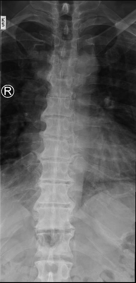
AP T-spineLabels for this film are coming.Image: IanWhyte. CC BY-SA 4.0. Source
Supine, knees flexed to reduce the kyphosis, MSP centered.
Central ray
Perpendicular
Centering
T7
Typical technique
75–85 kVp · 10–20 mAs · 40" SID
Image criteria
C7 through L1 included.
Spinous processes midline; no rotation.
Even density top to bottom (anode heel effect).
Lateral thoracic spine
CR: Perpendicular (10–15° cephalad if the spine isn't supported)
Position
Left lateral, arms forward at right angles, knees flexed; support under the waist so the spine is parallel to the table.
Central ray
Perpendicular (10–15° cephalad if the spine isn't supported)
Centering
T7
Typical technique
80–90 kVp · 25–50 mAs · 40–48" SID
Image criteria
Intervertebral foramina and disk spaces open.
Ribs and lung markings blurred by breathing.
Posterior ribs superimposed (no rotation).
Scoliosis series: PA (or AP) whole spine
Standing; Ferguson method for the block viewCR: Perpendicular, horizontal beam
Position
Standing (upright), weight evenly on both feet, arms at the sides, MSP centered to the long IR. PA (facing the IR) is preferred to reduce breast and thyroid dose; AP is used when the patient can't stand facing the IR. Gowned, with all metal removed from the trunk.
Central ray
Perpendicular, horizontal beam
Centering
MSP of the IR, positioned so the top is about 1–2 inches above C7 and the bottom about 1–2 inches below the iliac crests (about T10–T12 on a 14 × 36 or stitched long-length IR)
Typical technique
85–95 kVp · 15–30 mAs · 60–72" SID
Best shows
The lateral curve(s) of the thoracic and lumbar spine for Cobb angle measurement, vertebral rotation and the Risser stage of the iliac crests
Image criteria
Whole thoracic and lumbar spine included, from the lower C-spine (C7) through the iliac crests and SI joints.
Patient standing straight, not leaning; no rotation of the pelvis (iliac wings symmetric) unless caused by the curve.
Even density from the dense lumbar area to the thinner upper thorax (compensating filter or long-length processing).
Side marker and an upright (arrow) marker on the film.
Scoliosis series: lateral whole spine
CR: Perpendicular, horizontal beam
Position
Standing lateral, convex side of the curve toward the IR (usually left lateral), arms raised or crossed so the humeri clear the spine. MSP parallel to the IR, coronal plane perpendicular.
Central ray
Perpendicular, horizontal beam
Centering
Midcoronal plane, at about T10–T12 on a 14 × 36 or long-length IR; include C7 through the sacrum
Typical technique
90–100 kVp · 40–80 mAs · 60–72" SID
Best shows
Kyphosis and lordosis (sagittal balance), and spondylolisthesis, which often goes with scoliosis
Image criteria
C7 through the sacrum included.
Posterior ribs and the vertebral bodies close to superimposed (no rotation); some separation is expected from the curve itself.
Arms clear of the spine.
Even density from the upper T-spine through L5–S1.
Technique values are typical starting points; follow your department's technique chart.
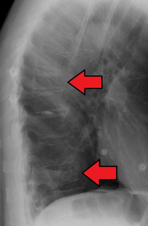
1. The arrows on this lateral chest radiograph point to which finding in the thoracic spine?
Image: James Heilman, MD. CC BY-SA 4.0.
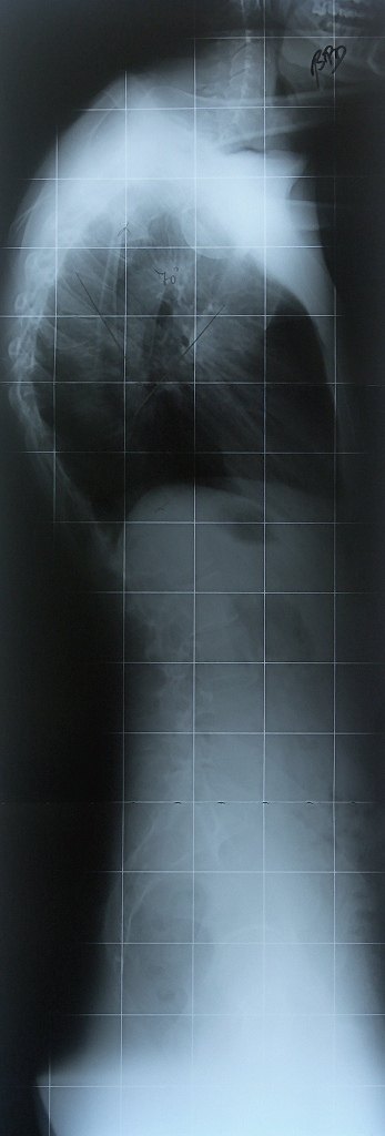
2. This erect lateral image from a scoliosis series has an angle drawn across the thoracic curve. Which abnormal curvature is being measured?
Image: ATarom. Public domain.
3. An AP thoracic spine shows the upper vertebrae too dark and the lower vertebrae too light, even after window adjustment, because of the large difference in part thickness. Which processing function can make both regions visible on one image?
4. Which projection of the thoracic spine BEST demonstrates the intervertebral foramina?
5. Image stitching software on a DR system is used MAINLY for which type of exam?