The shoulder girdle is the clavicle and scapula, joined to the humerus at the glenohumeral joint. A routine shoulder is AP external and internal rotation; trauma uses an AP in neutral plus a scapular Y.
Overview
Bones
Clavicle
Sternal end
Acromial end
Scapula
Acromion
Coracoid process
Glenoid cavity
Spine
Medial (vertebral) border
Lateral (axillary) border
Inferior angle
Proximal humerus
Head
Greater tubercle
Lesser tubercle
Surgical neck
Joints
Glenohumeral
Acromioclavicular (AC)
Sternoclavicular (SC)
Registry must-knows
AP external rotation: greater tubercle in profile laterally. AP internal rotation: lesser tubercle in profile medially.
Trauma: don't rotate the arm. Take an AP in neutral and a scapular Y.
On the scapular Y, the humeral head sits over the center of the Y. Anterior dislocation (the most common) puts it under the coracoid; posterior puts it under the acromion.
A Hill-Sachs defect is a compression fracture of the posterolateral humeral head; a Bankart lesion involves the anteroinferior glenoid rim.
Show 1 more
AC joints: erect, both sides on one image, with and without 5–8 lb weights hung from the wrists (not held in the hands), at 72 inches SID.
3D model
Loading 3D…
Radiographs 11
Pick a film, then a structure to circle it.
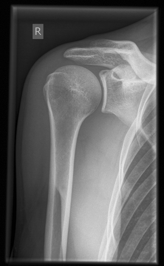
AP oblique shoulder (Grashey), right, arm internally rotatedImage: RSatUSZ / UniversitätsSpital Zürich. CC BY-SA 4.0. Source
Erect or supine, hand supinated, epicondyles parallel to the IR.
Central ray
Perpendicular
Centering
1 inch inferior to the coracoid process
Typical technique
70–75 kVp · 8–12 mAs · 40" SID
Image criteria
Greater tubercle in profile laterally.
Humeral head partly superimposes the glenoid.
Clavicle and upper scapula included.
AP shoulder, internal rotation
CR: Perpendicular
Position
Back of the hand against the thigh, epicondyles perpendicular to the IR.
Central ray
Perpendicular
Centering
1 inch inferior to the coracoid process
Typical technique
70–75 kVp · 8–12 mAs · 40" SID
Image criteria
Lesser tubercle in profile medially.
Greater tubercle superimposed over the humeral head.
Not used if a fracture is suspected.
AP oblique (glenoid cavity)
GrasheyCR: Perpendicular
Position
Rotate the body 35–45° toward the affected side so the scapula is parallel to the IR.
Central ray
Perpendicular
Centering
2 inches medial and 2 inches inferior to the superolateral border of the shoulder
Typical technique
70–75 kVp · 8–12 mAs · 40" SID
Best shows
Glenoid cavity in profile and an open glenohumeral joint
Image criteria
Glenoid cavity in profile.
Glenohumeral joint space open.
Scapular Y (PA oblique)
CR: Perpendicular
Position
Erect, affected side against the IR, body rotated 45–60° anterior oblique so the scapula is perpendicular to the IR.
Central ray
Perpendicular
Centering
Scapulohumeral joint
Typical technique
70–80 kVp · 12–20 mAs · 40" SID
Best shows
Anterior or posterior shoulder dislocation; scapular body fractures
Image criteria
Scapula in a true lateral: body seen on end, forming the Y with the acromion and coracoid.
Humeral head centered over the Y in a normal shoulder.
Ribs not superimposed on the scapular body.
Inferosuperior axial
LawrenceCR: 25–30° medially, horizontal, into the axilla
Position
Supine, arm abducted 90° and externally rotated if tolerated.
Central ray
25–30° medially, horizontal, into the axilla
Centering
Axilla / glenohumeral joint
Typical technique
70–75 kVp · 8–12 mAs · 40" SID
Best shows
Relationship of the humeral head to the glenoid; coracoid and lesser tubercle
Image criteria
Glenohumeral joint open.
Coracoid process and lesser tubercle in profile.
Acromion superimposed on the humeral head.
Tangential outlet
NeerCR: 10–15° caudad
Position
Scapular Y position (45–60° anterior oblique).
Central ray
10–15° caudad
Centering
Superior margin of the humeral head
Typical technique
70–80 kVp · 12–20 mAs · 40" SID
Best shows
The supraspinatus outlet (coracoacromial arch) for impingement
Image criteria
Humeral head below the outlet, not superimposed by the acromion.
Scapula in a true lateral.
AC joints (bilateral)
PearsonCR: Perpendicular
Position
Erect, both AC joints on one image; one exposure without and one with 5–8 lb weights hung from the wrists.
Central ray
Perpendicular
Centering
Midsagittal plane at the level of the AC joints
Typical technique
65–75 kVp · 8–12 mAs · 72" SID
Best shows
AC separation
Image criteria
Both AC joints on one image, at the same level.
Weights hung from the wrists, not held in the hands.
Technique values are typical starting points; follow your department's technique chart.
Labeled diagrams
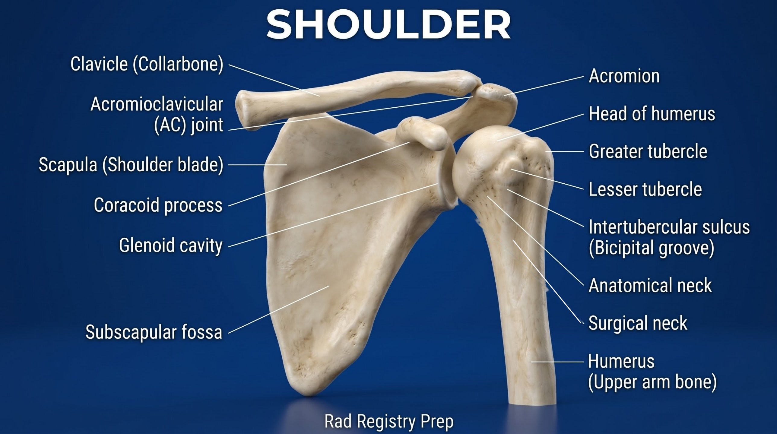
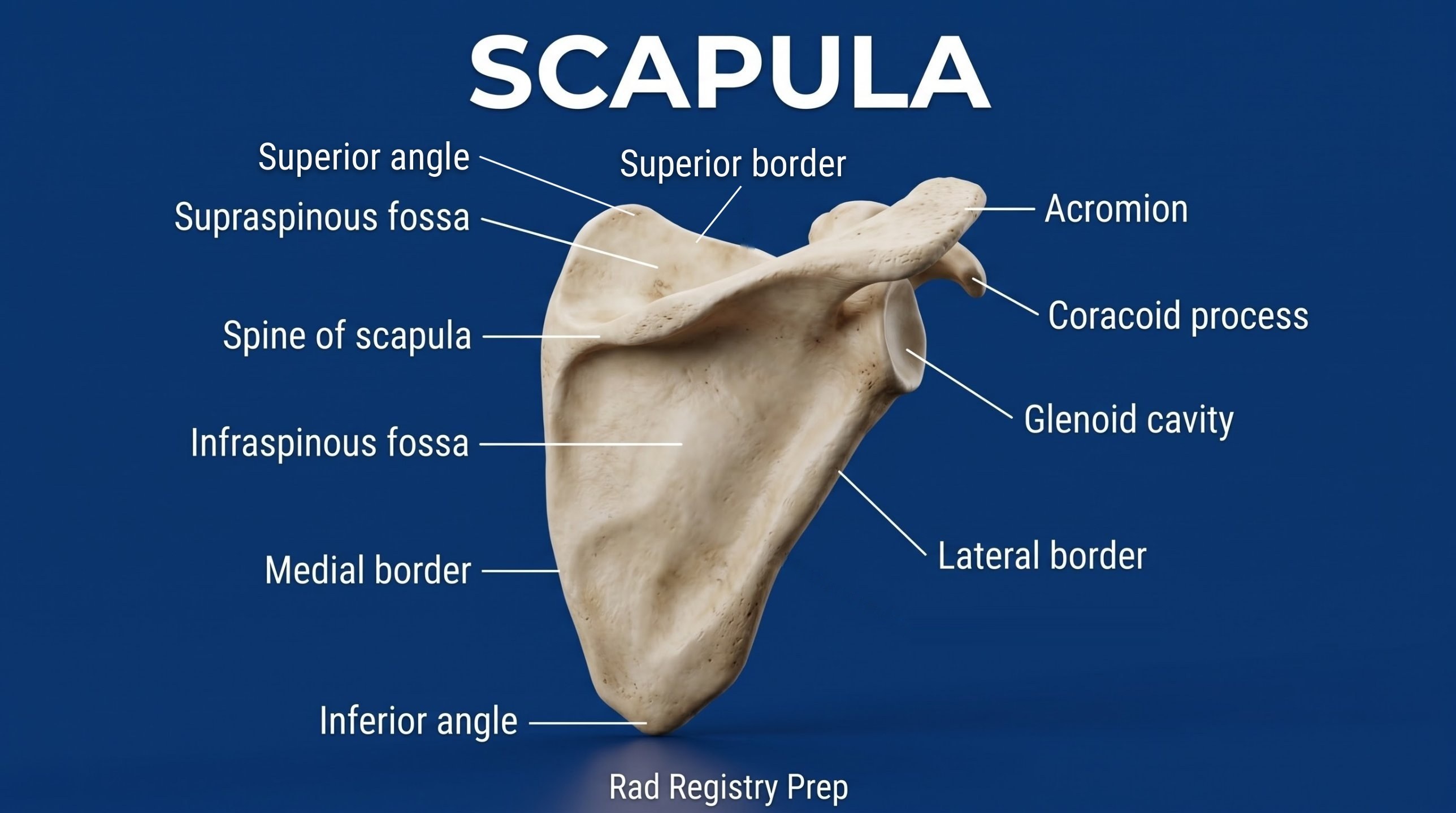
Shoulder (anterior view) · Diagram: Rad Registry Prep (original illustration).Scapula (posterior view) · Diagram: Rad Registry Prep (original illustration).
Quick check
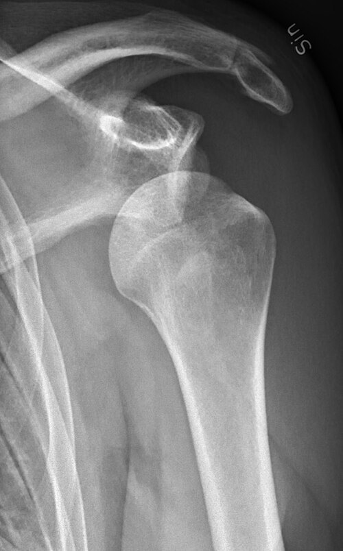
1. This AP shoulder radiograph of a trauma patient shows the humeral head displaced out of the glenoid cavity. To show whether the dislocation is anterior or posterior without abducting or rotating the arm, which projection should the radiographer perform?
Image: Mikael Häggström, M.D. CC0 1.0.
2. For the tangential outlet projection (Neer method) to evaluate shoulder impingement, the patient is positioned as for a scapular Y. How is the CR directed?
3. A patient has suspected shoulder impingement. Which projection demonstrates the supraspinatus outlet?
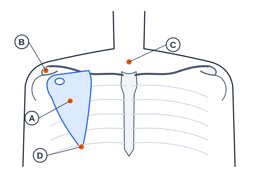
4. The AP schematic shows the patient's right scapula (shaded) with the arm abducted 90 degrees; the small oval marks the coracoid process. For an AP projection of the scapula, at which labeled point is the CR directed?
Original diagram: Rad Registry Prep.
5. A patient has pain and swelling at the left sternoclavicular joint. Which position BEST shows the left SC joint?