RAD Registry Prep logo

Anatomy & Positioning Atlas

Lumbar Spine

The lumbar spine has 5 large vertebrae above the sacrum. A routine L-spine is an AP, obliques, a lateral and a spot lateral of L5–S1.

Overview

Bones

Lumbar vertebrae
  1. Body
  2. Pedicles
  3. Laminae
  4. Pars interarticularis
  5. Superior and inferior articular processes
  6. Transverse processes
  7. Spinous process
Below
  1. Sacrum
  2. Sacral promontory
  3. Coccyx
  4. SI joints

Scottie dog on the oblique: nose = transverse process, eye = pedicle, ear = superior articular process, neck = pars interarticularis, front leg = inferior articular process. A collar on the neck is spondylolysis.

Registry must-knows

  • AP: knees flexed to flatten the lumbar curve and open the disk spaces.
  • Obliques (45°): posterior obliques (RPO/LPO) show the zygapophyseal joints closest to the IR; anterior obliques show those farthest.
  • L5–S1 spot lateral: CR to L5–S1, about 1.5 inches below the iliac crest and 2 inches posterior to the ASIS; angle 5–8° caudad if the waist isn't supported.
  • Spondylolisthesis (forward slip of a vertebra) shows best on the lateral; spondylolysis (pars defect) on the obliques.
Show 1 more
  • Iliac crest = L4–L5; ASIS = S1–S2.

3D model

Loading 3D…

Radiographs 4

Pick a film, then a structure to circle it.

Projections 4

Tap a projection for positioning, central ray and image criteria. See every exam in the Positioning Guide ›

AP lumbar spine

CR: Perpendicular
Position
Supine, knees flexed, MSP centered.
Central ray
Perpendicular
Centering
Iliac crest (L4–L5) for a 14 × 17 IR; L3 (about 1.5 inches above the crest) for smaller IRs
Typical technique
80–90 kVp · 20–40 mAs · 40" SID

Image criteria

  • T12 through the sacrum included; psoas margins seen.
  • Spinous processes midline; no rotation.
  • Intervertebral disk spaces open.

AP oblique (posterior oblique)

RPO / LPO
CR: Perpendicular
Position
Body rotated 45° from supine (posterior oblique).
Central ray
Perpendicular
Centering
L3, about 2 inches medial to the elevated ASIS and 1.5 inches above the iliac crest
Typical technique
80–90 kVp · 20–40 mAs · 40" SID
Best shows
Zygapophyseal joints closest to the IR (the 'Scottie dog')

Image criteria

  • Zygapophyseal joints open.
  • Pedicle (eye) near the center of the body.
  • Scottie dog outlines seen.

Lateral lumbar spine

CR: Perpendicular (5–8° caudad if the waist isn't supported)
Position
Left lateral, knees and hips flexed and stacked, support under the waist so the spine is parallel to the table.
Central ray
Perpendicular (5–8° caudad if the waist isn't supported)
Centering
L4 at the iliac crest (L3 for smaller IRs)
Typical technique
85–95 kVp · 40–80 mAs · 40" SID

Image criteria

  • T12 through the sacrum included.
  • Intervertebral foramina and disk spaces open.
  • Posterior vertebral bodies superimposed (no rotation).

L5–S1 spot lateral

CR: Perpendicular (5–8° caudad if the waist isn't supported)
Position
Left lateral, as for the lateral L-spine.
Central ray
Perpendicular (5–8° caudad if the waist isn't supported)
Centering
1.5 inches below the iliac crest, 2 inches posterior to the ASIS
Typical technique
90–100 kVp · 60–100 mAs · 40" SID

Image criteria

  • L5–S1 joint space open.
  • No rotation: greater sciatic notches superimposed.

Technique values are typical starting points; follow your department's technique chart.

Labeled diagrams

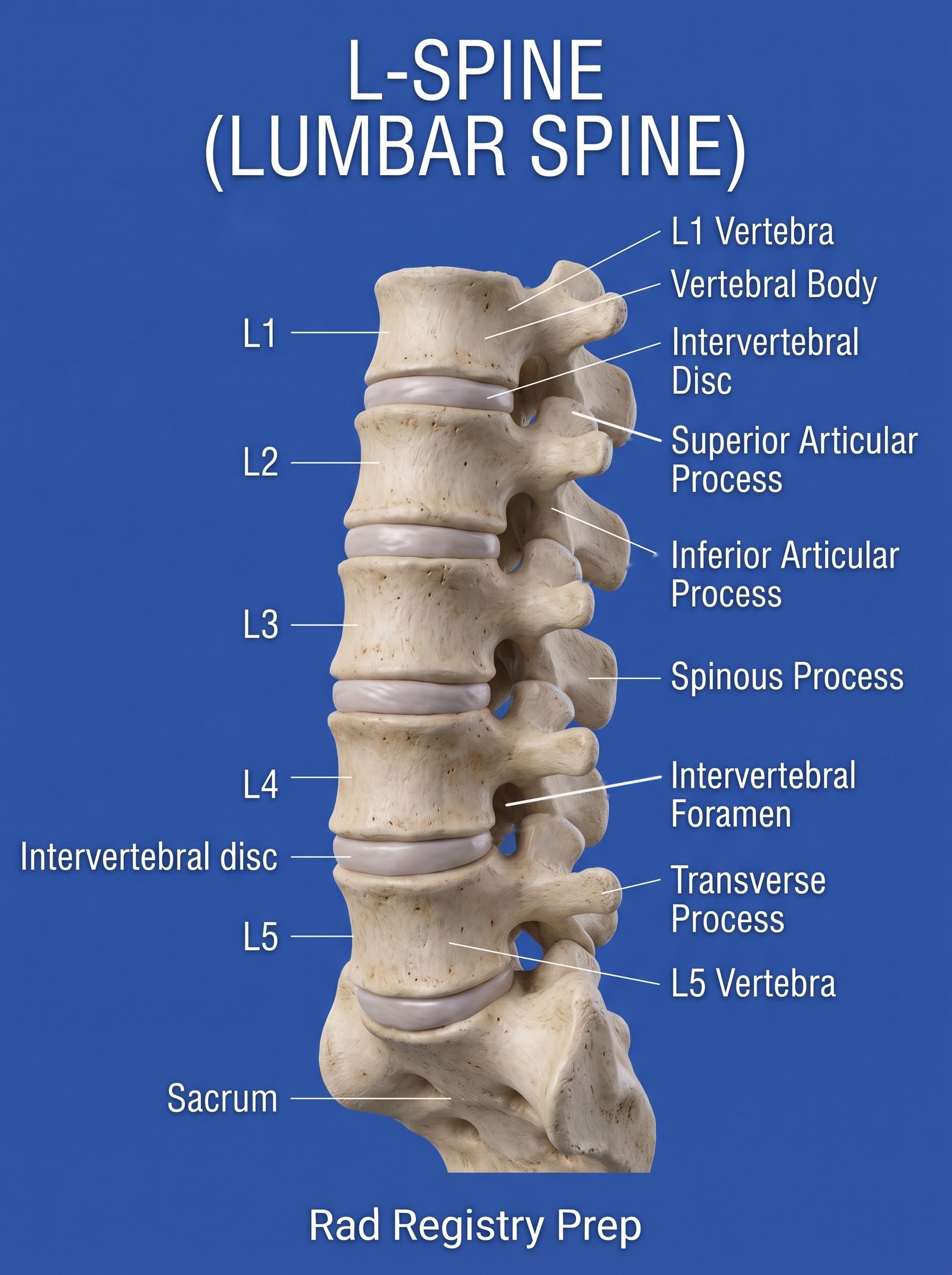
Labeled lumbar spine diagram (lateral): L1, L2, L3, L4, L5, L1 vertebra, Vertebral body, Intervertebral disc, Superior articular process, Inferior articular process, Spinous process, Intervertebral foramen, Transverse process, L5 vertebra, Sacrum.
Lumbar Spine (lateral view) · Diagram: Rad Registry Prep (original illustration).

Quick check

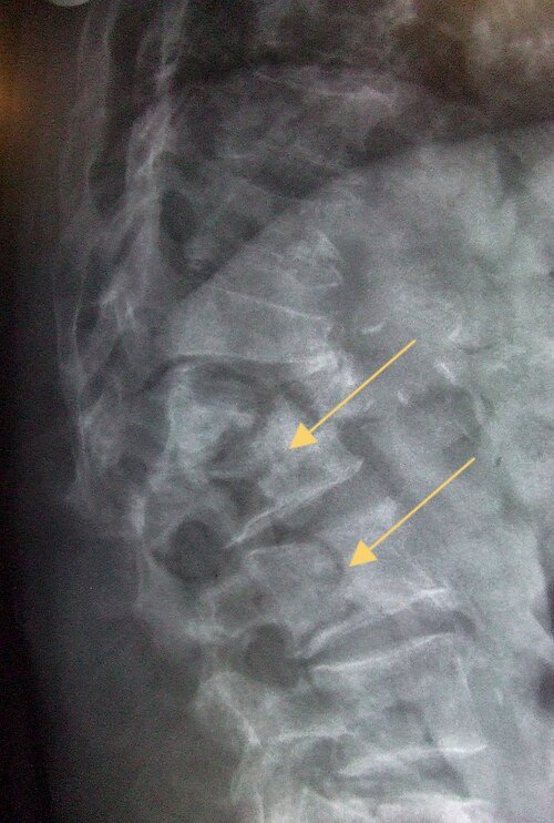
1. A 78-year-old woman reports back pain after lifting a laundry basket. The arrows on this lateral spine radiograph mark vertebral bodies with anterior height loss (compression fractures). Which underlying condition MOST often causes this type of fracture after minimal trauma?

Image: Glitzy queen00 / Wikimedia Commons. Public domain.

2. Which radiographic examination typically delivers the HIGHEST dose to the patient's gonads?

3. For the AP axial projection of the coccyx, how is the CR directed?

4. A 30-year-old outpatient having AP and lateral lumbar spine radiographs asks for a lead shield over her pelvis. The department follows current AAPM guidance. Which response is MOST appropriate?

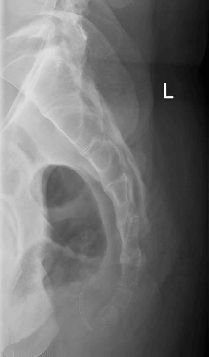
5. This lateral sacrum and coccyx was taken for chronic tailbone pain. Which centering point is used for a lateral coccyx?

Image: Algain A, Karami M, Kinsara A, Qoqandi MA, Alsulimany H, Cureus 2024, via PubMed Central. CC BY 4.0.

Full procedures quiz →

← Return to Anatomy Atlas