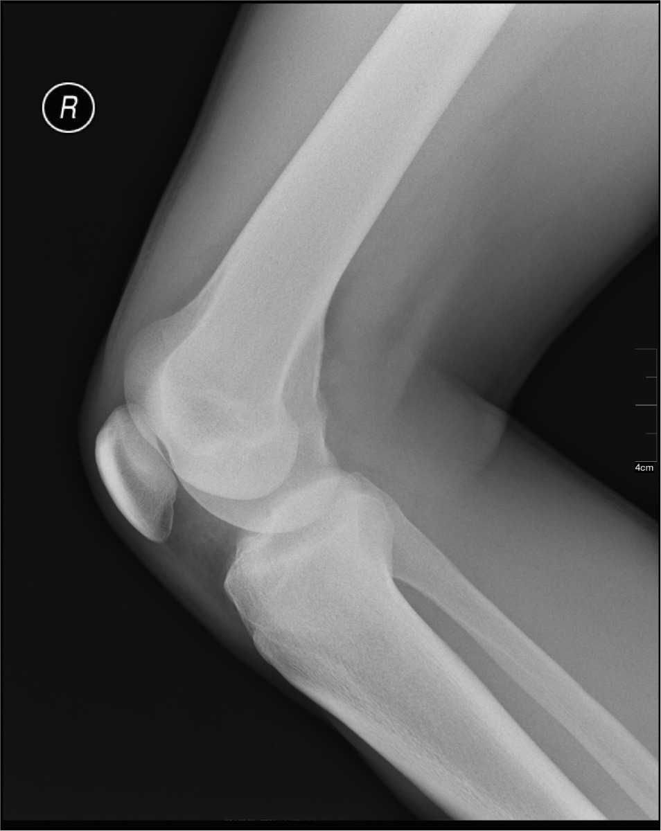
1. On a lateral knee radiograph, the femoral condyles are offset from each other in the anterior-posterior direction instead of being superimposed. Which positioning error is MOST likely responsible?
Knee
The knee joins the femur, tibia and patella. A routine knee is an AP, a lateral and an oblique; special views add the intercondylar fossa (tunnel) and the patella.
Overview
Bones
Distal femur
- Medial condyle
- Lateral condyle
- Medial epicondyle
- Lateral epicondyle
- Intercondylar fossa
Proximal tibia
- Medial condyle
- Lateral condyle
- Tibial plateau
- Intercondylar eminence
- Tibial tuberosity
Others
- Patella (largest sesamoid)
- Fibular head
AP knee CR angle by ASIS-to-tabletop distance: under 19 cm, 3–5° caudad; 19–24 cm, 0°; over 24 cm, 3–5° cephalad.
Registry must-knows
- Lateral knee: flex only 20–30° (more tightens the quadriceps and closes the patellofemoral space) and angle the CR 5–7° cephalad so the condyles superimpose.
- AP medial oblique (45° internal rotation) opens the proximal tibiofibular joint and frees the fibular head; the lateral oblique superimposes them.
- Weight-bearing PA or AP knees show the joint spaces narrowing in arthritis.
- Intercondylar fossa: Camp-Coventry (prone, CR 40–50° caudad to match flexion), Holmblad (kneeling, CR perpendicular to the lower leg).
Show 1 more
- Patella tangential: Settegast (knee flexed 90°+, CR 15–20° to the joint) and Merchant (knees flexed 40°, CR 30° caudad).
3D model
Loading 3D…
Radiographs 6
Pick a film, then a structure to circle it.
Projections 6
Tap a projection for positioning, central ray and image criteria. See every exam in the Positioning Guide ›
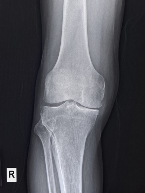
AP knee
CR: 0° to 5° cephalad or caudad depending on the ASIS-to-tabletop measurement
- Position
- Supine, leg extended and rotated 3–5° internally so the epicondyles are parallel to the IR.
- Central ray
- 0° to 5° cephalad or caudad depending on the ASIS-to-tabletop measurement
- Centering
- 1/2 inch distal to the apex of the patella
- Typical technique
- 70–75 kVp · 6–10 mAs · 40" SID
Image criteria
- Femorotibial joint space open.
- No rotation: symmetric femoral and tibial condyles; fibular head about half superimposed by the tibia.
- Patella slightly medial of midline.
Lateral knee (mediolateral)
CR: 5–7° cephalad
- Position
- Lateral recumbent on the affected side, knee flexed 20–30°, opposite leg behind.
- Central ray
- 5–7° cephalad
- Centering
- 1 inch distal to the medial epicondyle
- Typical technique
- 70–75 kVp · 6–10 mAs · 40" SID
Image criteria
- Femoral condyles superimposed.
- Patellofemoral joint space open; patella in profile.
- Fibular head slightly superimposed by the tibia.
AP oblique, medial (internal) rotation
CR: Same angle as the AP
- Position
- Supine, leg rotated 45° internally.
- Central ray
- Same angle as the AP
- Centering
- 1/2 inch distal to the apex of the patella
- Typical technique
- 70–75 kVp · 6–10 mAs · 40" SID
- Best shows
- Proximal tibiofibular joint and fibular head free of superimposition; lateral condyles
Image criteria
- Fibular head and neck free of the tibia.
- Tibiofibular joint open.
- Lateral femoral and tibial condyles in profile.
AP oblique, lateral (external) rotation
CR: Same angle as the AP
- Position
- Supine, leg rotated 45° externally.
- Central ray
- Same angle as the AP
- Centering
- 1/2 inch distal to the apex of the patella
- Typical technique
- 70–75 kVp · 6–10 mAs · 40" SID
- Best shows
- Medial condyles in profile; fibula superimposed by the tibia
Image criteria
- Medial femoral and tibial condyles in profile.
- Fibular head superimposed by the tibia.
PA axial intercondylar fossa
Camp-CoventryCR: 40–50° caudad, matching the knee flexion (perpendicular to the lower leg)
- Position
- Prone, knee flexed 40–50° on a support.
- Central ray
- 40–50° caudad, matching the knee flexion (perpendicular to the lower leg)
- Centering
- Popliteal crease
- Typical technique
- 70–75 kVp · 8–12 mAs · 40" SID
- Best shows
- Intercondylar fossa and the intercondylar eminence (loose bodies, osteochondritis)
Image criteria
- Intercondylar fossa open, in profile.
- No rotation: symmetric posterior condyles.
Tangential patella
Settegast / MerchantCR: Settegast: 15–20° to the patellofemoral joint. Merchant: 30° caudad from horizontal.
- Position
- Settegast: prone or seated, knee flexed at least 90°. Merchant: supine, both knees flexed 40° over the end of the table.
- Central ray
- Settegast: 15–20° to the patellofemoral joint. Merchant: 30° caudad from horizontal.
- Centering
- Patellofemoral joint
- Typical technique
- 65–75 kVp · 6–12 mAs · 40" SID
- Best shows
- Patellofemoral joint space and vertical patellar fractures
Image criteria
- Patella seen in profile above the femoral condyles.
- Patellofemoral joint space open.
Technique values are typical starting points; follow your department's technique chart.
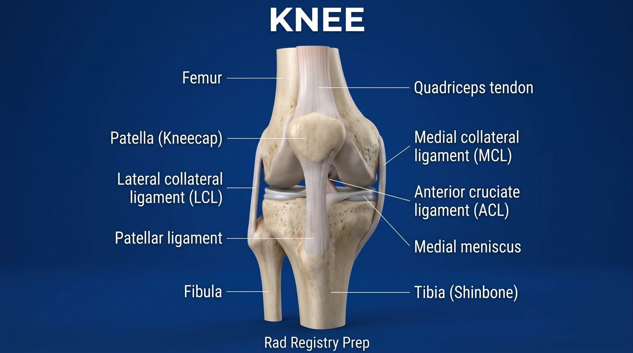
Labeled diagrams
Quick check
2. The patella is the largest sesamoid bone in the body. Within the tendon of which muscle does it develop?
3. For the PA axial (Holmblad) projection of the intercondylar fossa, the knee is flexed approximately how many degrees?
4. For a tangential projection of the patella, the patient lies prone on the table, the knee is flexed about 55 degrees, and a strap is looped around the foot. Which method is being performed?
5. This lateral knee is flexed far more than usual, pulling the patella down into the femoral groove. Why is a lateral knee normally flexed only 20–30°?