The hip is the ball-and-socket joint between the femoral head and the acetabulum. A routine hip is an AP and a lateral; the frog-leg lateral is never used when a fracture is suspected, so trauma gets a cross-table axiolateral instead.
Overview
Bones
Proximal femur
Head
Fovea capitis
Neck
Greater trochanter
Lesser trochanter
Intertrochanteric crest
Hip bone
Acetabulum
ASIS
Ischial tuberosity
Superior pubic ramus
Locate the femoral neck: draw a line between the ASIS and the pubic symphysis; the neck is about 2.5 inches distal to its midpoint, on a perpendicular.
Registry must-knows
Internal rotation of 15–20° puts the femoral neck parallel to the IR. A shortened, externally rotated leg suggests a hip fracture: don't rotate it.
Frog-leg (modified Cleaves): hip abducted 40–45° — contraindicated in trauma.
Cross-table axiolateral (Danelius-Miller): unaffected leg raised out of the way, horizontal CR perpendicular to the femoral neck.
Bilateral hip trauma: the modified axiolateral (Clements-Nakayama) angles the CR 15° posteriorly instead of raising the other leg.
Show 1 more
Slipped capital femoral epiphysis (SCFE) shows best on the frog-leg lateral in adolescents.
3D model
Loading 3D…
Radiographs 5
Pick a film, then a structure to circle it.
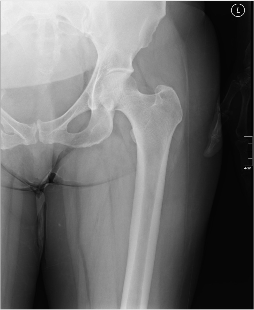
AP proximal femur and hipImage: Nevit Dilmen, via Wikimedia Commons. CC BY-SA 3.0. Source
Semi-supine, body rotated 45°. Obturator oblique: affected side UP (raised). Iliac oblique: affected side DOWN, against the table. Both are usually taken.
Central ray
Perpendicular
Centering
Affected side up: 2 inches distal and 2 inches medial to the upside ASIS. Affected side down: 2 inches distal to the downside ASIS
Typical technique
80–85 kVp · 20–30 mAs · 40" SID
Best shows
Obturator oblique: the anterior column (iliopubic) and posterior rim of the acetabulum, with the obturator foramen open. Iliac oblique: the posterior column (ilioischial), the anterior rim and the iliac wing in profile.
Image criteria
Acetabulum and femoral head in the center of the image.
Iliac oblique: iliac wing open and in profile, obturator foramen closed.
AP hip
CR: Perpendicular
Position
Supine, leg rotated 15–20° internally (not in trauma).
Central ray
Perpendicular
Centering
Femoral neck (about 2.5 inches distal to the midpoint of the ASIS–symphysis line)
Typical technique
80–85 kVp · 15–30 mAs · 40" SID
Image criteria
Femoral neck in full length, without foreshortening.
Greater trochanter in profile; lesser trochanter barely seen.
Acetabulum, femoral head and proximal third of the femur included.
Lateral hip, frog-leg
Modified CleavesCR: Perpendicular
Position
Supine, knee flexed and the thigh abducted 40–45° (or as tolerated). Not used in trauma.
Central ray
Perpendicular
Centering
Femoral neck
Typical technique
80–85 kVp · 15–40 mAs · 40" SID
Image criteria
Femoral neck seen laterally, superimposed by the greater trochanter.
Lesser trochanter on the medial side.
Axiolateral hip
Danelius-MillerCR: Horizontal, perpendicular to the femoral neck and IR
Position
Supine, unaffected leg flexed and raised out of the way; IR in the crease above the iliac crest, parallel to the femoral neck.
Central ray
Horizontal, perpendicular to the femoral neck and IR
Centering
Femoral neck
Typical technique
80–90 kVp · 32–80 mAs · 40" SID
Best shows
A lateral of the proximal femur without moving the injured leg (trauma)
Image criteria
Femoral head, neck and trochanters in profile.
Unaffected leg out of the field; no grid lines.
Technique values are typical starting points; follow your department's technique chart.
Labeled diagrams
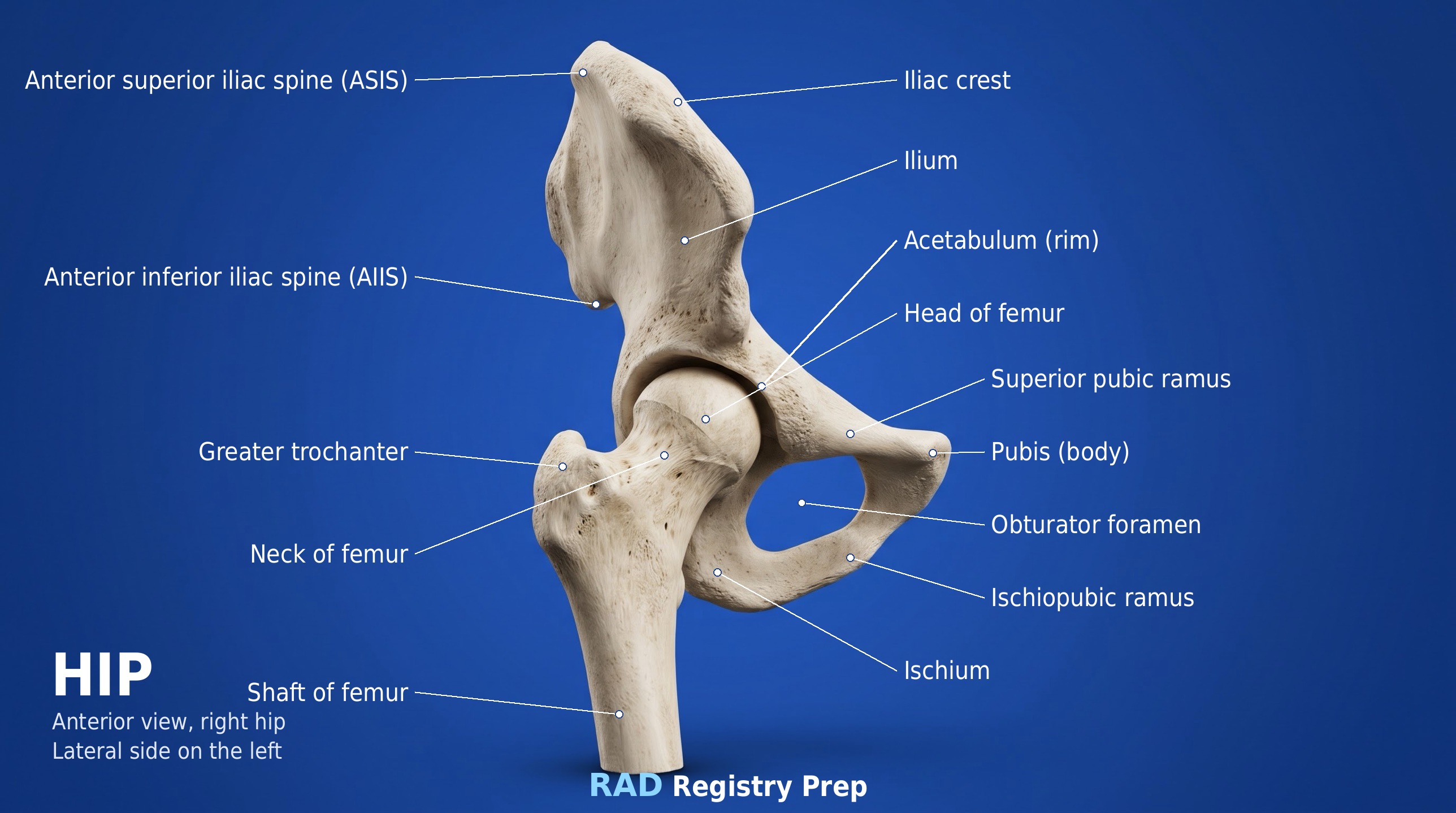
Hip (anterior, right hip (lateral side on the left) view) · Diagram: Rad Registry Prep (original illustration).
Quick check
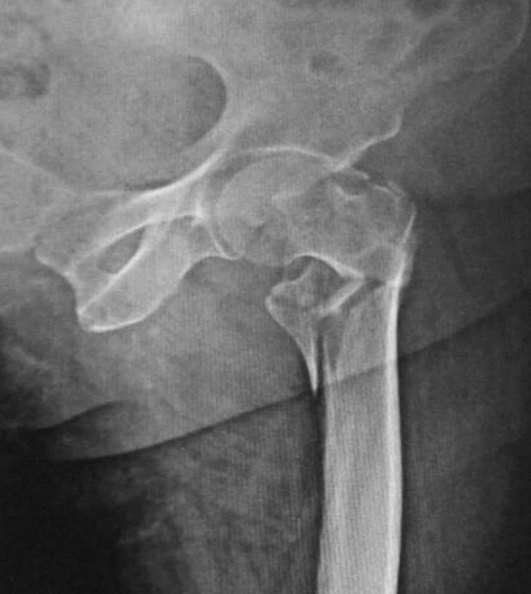
1. This AP hip radiograph of a patient in the emergency department shows a displaced, comminuted proximal femur fracture. The patient can flex and raise the unaffected leg. Which lateral projection should the radiographer perform?
Image: Memon, Patel & Juva. CC BY 4.0.
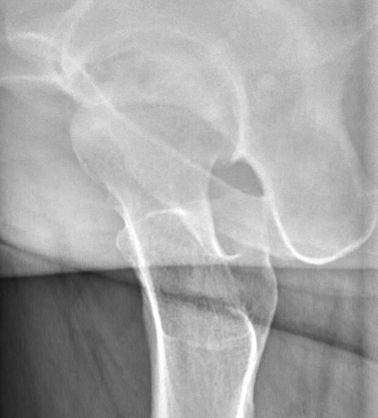
2. A trauma patient needs a lateral of this hip but has bilateral hip injuries and cannot flex or elevate the opposite leg. Which projection should replace the Danelius-Miller?
Image: Mikael Häggström, M.D. CC0 1.0.
3. A patient has a suspected left femoral neck fracture. The right hip cannot be flexed or elevated because of a recent hip replacement. Which lateral projection of the left hip is MOST appropriate?
4. A patient with a suspected right hip fracture arrives with the right foot externally rotated. For the AP pelvis, what should the radiographer do?
5. A non-trauma AP proximal femur and hip is reviewed. The lesser trochanter is seen fully in profile along the medial side of the femur, and the femoral neck is foreshortened. What caused this appearance?