The ankle (talocrural) joint is the mortise formed by the tibia and fibula around the talus. Routine ankle imaging is an AP, a mortise and a lateral; some departments add a 45° oblique.
Overview
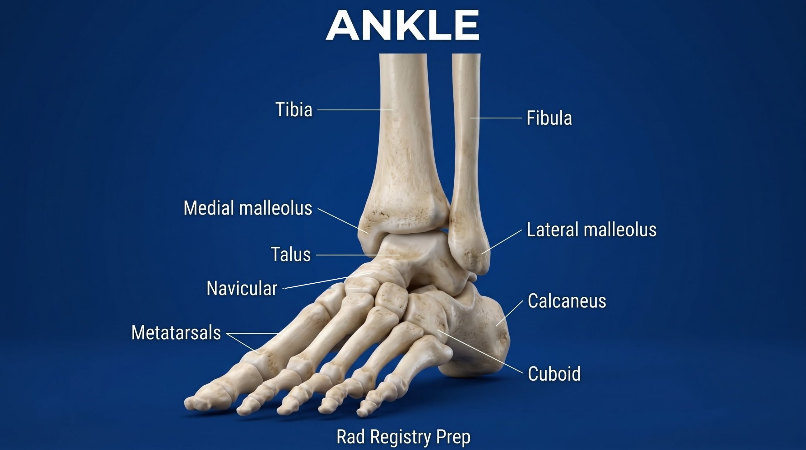
Bones
Mortise
Distal tibia (plafond)
Medial malleolus
Lateral malleolus (fibula)
Talus
Trochlea (dome)
Neck
Head
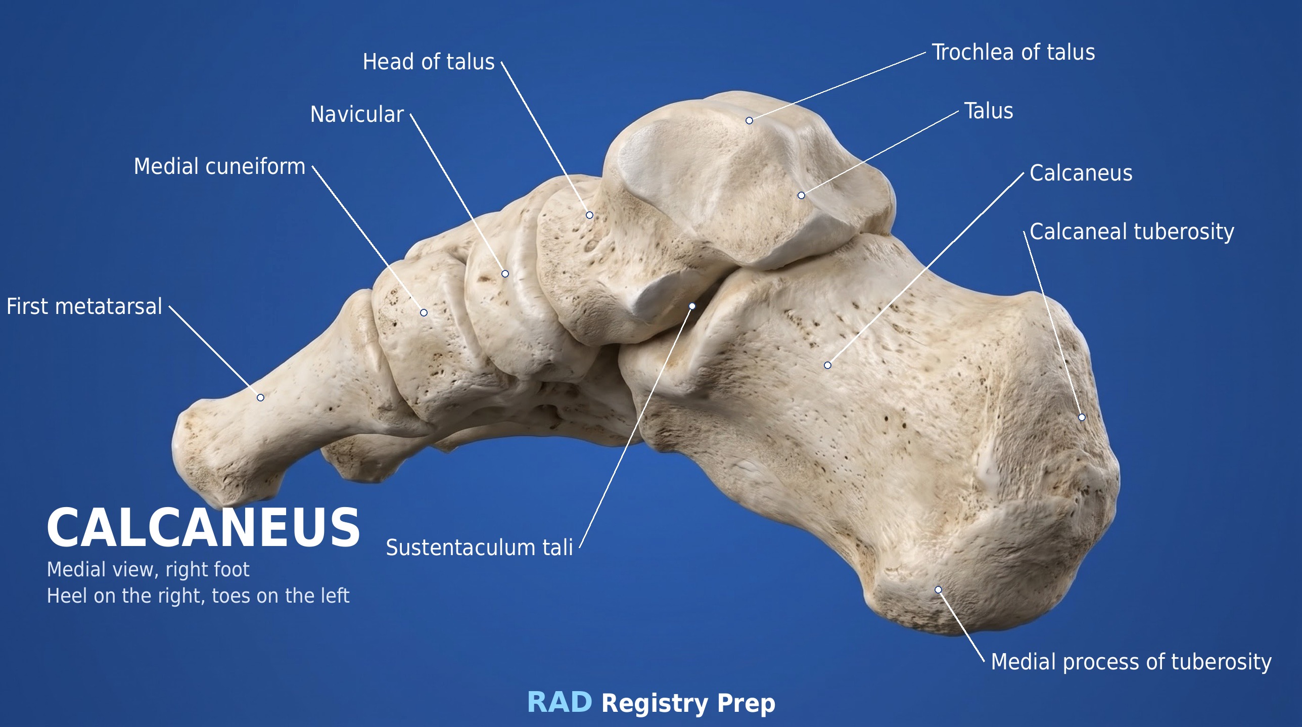
Calcaneus
Tuberosity
Sustentaculum tali
Mortise: rotate the leg in 15–20° until the line between the malleoli is parallel to the IR. Then the whole joint space is open.
Registry must-knows
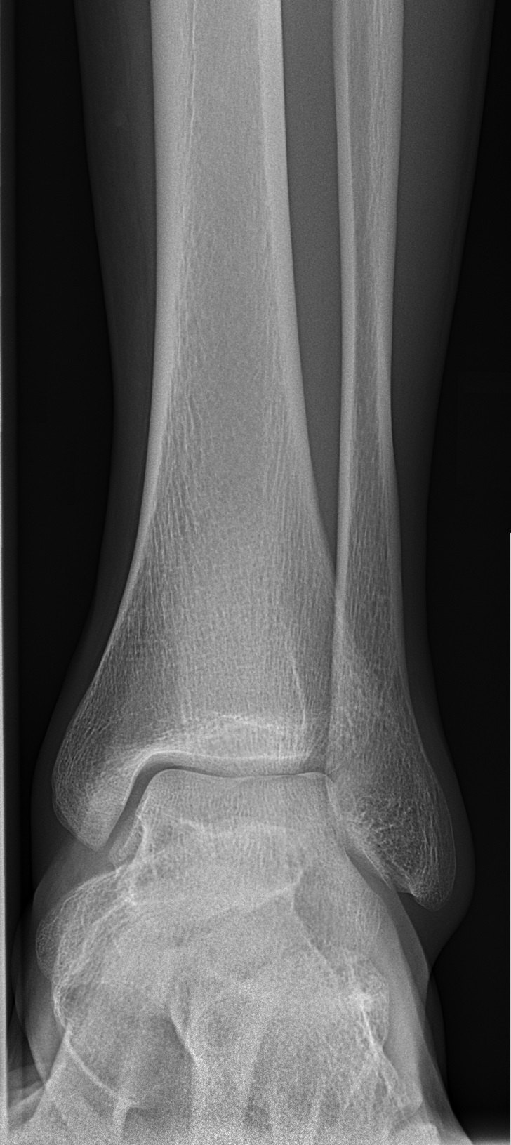
AP ankle: the lateral mortise is closed because the fibula overlaps the talus — that's normal on the AP.
Mortise view: the entire mortise open, including the medial and lateral clear spaces, with no overlap of the talus and malleoli.
Lateral ankle: the fibula lies over the posterior half of the tibia; include the base of the 5th metatarsal to catch an avulsion.
Stress views (inversion/eversion) are done by a physician or qualified person, never forced by the radiographer.
Show 1 more
Trimalleolar fracture: medial and lateral malleoli plus the posterior lip of the tibia.
3D model
Loading 3D…
Radiographs 3
Pick a film, then a structure to circle it.
AP ankleImage: Mikael Häggström, M.D., via Wikimedia Commons. CC0 (public domain dedication). Source
Supine, leg extended, foot dorsiflexed with the toes straight up.
Central ray
Perpendicular
Centering
Midway between the malleoli
Typical technique
60–65 kVp · 3–5 mAs · 40" SID
Image criteria
Distal tibia and fibula, malleoli, talus and proximal metatarsals included.
Medial mortise open; lateral mortise closed (fibula overlaps the talus).
Tibiotalar joint open.
AP mortise
CR: Perpendicular
Position
From the AP, rotate the leg and foot 15–20° internally until the intermalleolar line is parallel to the IR.
Central ray
Perpendicular
Centering
Midway between the malleoli
Typical technique
60–65 kVp · 3–5 mAs · 40" SID
Best shows
The entire ankle mortise
Image criteria
Entire mortise open: medial, superior and lateral joint spaces equal.
Talus free of the malleoli.
Malleoli in profile.
AP oblique, 45° medial rotation
CR: Perpendicular
Position
From the AP, rotate the leg and foot 45° internally.
Central ray
Perpendicular
Centering
Midway between the malleoli
Typical technique
60–65 kVp · 3–5 mAs · 40" SID
Best shows
Distal tibiofibular joint and lateral malleolus
Image criteria
Distal tibiofibular joint open.
Lateral malleolus free of the talus.
Lateral ankle (mediolateral)
CR: Perpendicular
Position
Lateral recumbent on the affected side, foot dorsiflexed, true lateral.
Central ray
Perpendicular
Centering
Medial malleolus
Typical technique
60–65 kVp · 3–5 mAs · 40" SID
Image criteria
Fibula over the posterior half of the tibia.
Talar dome and tibiotalar joint open.
Base of the 5th metatarsal included.
Technique values are typical starting points; follow your department's technique chart.
Labeled diagrams
Ankle (anterior view) · Diagram: Rad Registry Prep (original illustration).Calcaneus (medial, right foot (heel on the right) view) · Diagram: Rad Registry Prep (original illustration).
Quick check
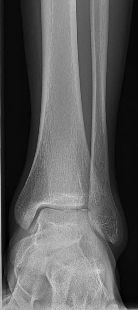
1. On this AP ankle, the lateral mortise is closed and the distal fibula overlaps the talus. Which change produces an AP mortise projection?
Image: Mikael Häggström, M.D. CC0 1.0.
2. For the AP mortise projection of the ankle, the leg and foot are rotated 15° to 20° medially. Which anatomic relationship makes this rotation necessary?
3. Inversion and eversion stress AP projections of the ankle are performed to evaluate:
4. A radiographer intends to obtain an AP mortise projection of the ankle. The image shows the distal tibiofibular joint wide open and the lateral malleolus completely free of the tibia. What does this indicate?
5. Inversion and eversion stress AP projections of the ankle are ordered. Who should apply the stress to the foot during the exposures?