
Patient Care · Assistance, monitoring and emergencies2 min read5-question check
Trauma and Emergency Response
Move the tube and receptor, not the patient, and keep immobilization on until a physician clears the spine.
The rule
A trauma patient must be imaged without making any injury worse. Leave cervical collars, backboards, and splints in place until a physician has cleared the spine or ordered them removed. Do not rotate an injured limb or roll the patient into a routine position. Instead, adapt: move the x-ray tube and the receptor around the patient. Speed matters, but so does getting the image right the first time.
Trauma workflow
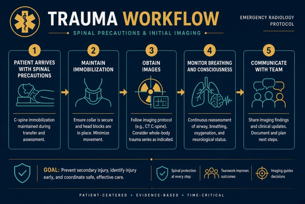
How it works
Adapted trauma imaging still follows the core rules. Obtain two projections at 90 degrees to each other, because a single view can hide displacement. Include both joints for a long bone. For the cervical spine, a cross-table lateral with a horizontal beam is taken first, with the collar on, and a physician reviews it before any other projections; all seven cervical vertebrae must be seen. Pulling the arms down to lower the shoulders helps show C7, but only with physician approval. A horizontal beam also shows air-fluid levels and free air when the patient cannot stand. If the patient must be moved, use a log roll with enough staff to keep the spine aligned.
On the job
Watch the patient as closely as the image. Signs of shock include falling blood pressure, a rapid weak pulse, pale cool clammy skin, restlessness, and anxiety. With a head injury, watch for a changing level of consciousness, unequal pupils, vomiting, or confusion. Never leave the patient alone, use standard precautions with blood and body fluids, and tell the trauma team at once about any change. Collimate tightly and keep your distance during every exposure.
Clinical Example
A motorcyclist arrives on a backboard with a cervical collar and a deformed forearm. You take a cross-table lateral cervical spine without moving the head, then image the forearm by moving the tube and receptor instead of rotating the arm. Midway through, the patient becomes confused and pale, so you stop and alert the team.
Common Mistakes
- Removing a cervical collar for a cleaner image before the spine is cleared.
- Rotating an injured limb into a routine position instead of angling the tube and receptor.
- Accepting a lateral cervical spine that does not show C7.
- Assuming a patient who arrived talking will stay stable.
Why It Matters
Moving an unstable spine or fracture can cause paralysis or further injury, and missed shock can be fatal. The registry tests who may remove immobilization, horizontal-beam technique, and recognizing shock.
Key Relationships
- Immobilization stays on until a physician clears it
- Move the tube and receptor, not the patient
- Two projections 90 degrees apart; both joints for long bones
- Lateral cervical spine must show all seven vertebrae
- Falling blood pressure + rapid weak pulse + cool clammy skin → shock