
Procedures · Head, spine and pelvis3 min read5-question check
Thoracic spine film check
AP on expiration with the cathode at the feet, a lateral with breathing technique to blur the ribs, and a swimmer's when the shoulders hide the top.
The AP
The AP thoracic spine runs from C7 to L1, centered at T7, about 3 to 4 inches below the jugular notch. The upper thoracic spine is thin and the lower part sits behind the dense heart and diaphragm, so it is easy to end up with one end too dark and the other too light. Two things even it out: put the cathode end of the tube toward the feet, so the stronger side of the beam covers the thicker lower spine, and expose on expiration, which leaves less air in the lungs, so the upper thoracic spine is not overexposed next to the denser lower spine. On a straight AP, the spinous processes sit in the midline and the sternoclavicular joints are the same distance from the spine.
The lateral
The lateral thoracic spine is usually a left lateral with the arms raised 90 degrees to the body, so the humeri and scapulae move out of the way. Put a support under the waist if needed so the spine is parallel to the table, and center at T7. On a good lateral the intervertebral disc spaces are open and the back edges of the vertebral bodies line up. Doubled posterior ribs or vertebral body edges mean rotation; stack the shoulders and the hips. A lead blocker behind the patient cuts the scatter that fogs the lateral.
Breathing technique
Breathing technique, also called orthostatic technique, uses a long exposure time, at least 3 to 4 seconds at a low mA, while the patient breathes slowly and gently. The ribs and lung markings move during the exposure and blur out, while the spine, which does not move, stays sharp. Tell the patient to breathe gently, not deeply, and to keep the rest of the body still. If the spine itself is blurred, the patient moved, not just breathed.
The top of the T-spine
The upper two or three thoracic vertebrae are usually hidden behind the shoulders on the lateral. To show them, take a swimmer's lateral, the cervicothoracic lateral: raise the arm closest to the IR over the head and push the other shoulder down, so the beam passes between the humeral heads.
Read the films
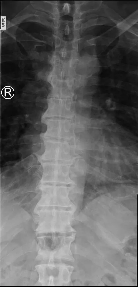
KeeperAP thoracic spine: a keeper
Look for: The spinous processes are in the midline and the whole thoracic spine is included, with even density from the top to the bottom.
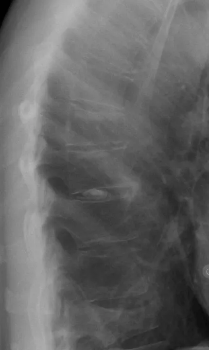
KeeperLateral thoracic spine
Look for: The vertebral bodies are in profile and the ribs and lung markings are soft behind them. This patient has a compression fracture of T12, where the front of the body is shorter than the back.
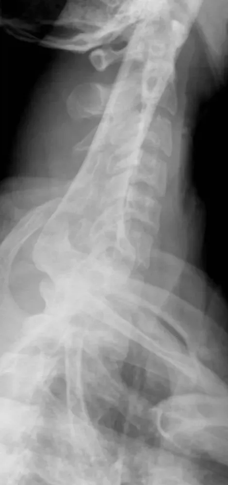
KeeperSwimmer's lateral: a keeper
Look for: C7 and the upper thoracic vertebrae show between the humeral heads, the part a routine lateral hides.
Clinical Example
Your lateral thoracic spine shows sharp ribs crossing the vertebral bodies and making them hard to read. You repeat with breathing technique: a 4-second exposure at low mA while the patient breathes slowly. The ribs blur out and the vertebral bodies are clear.
Common Mistakes
- Putting the anode toward the feet, which leaves the lower thoracic spine underexposed.
- Telling the patient to breathe deeply during breathing technique; gentle breaths blur the ribs without moving the spine.
- Leaving the arms down on the lateral.
- Expecting the upper thoracic spine to show on a routine lateral; add a swimmer's.
Why It Matters
Compression fractures of the thoracic spine are common in older patients and in trauma. The registry tests the anode heel effect, breathing technique and the swimmer's lateral.
Key Relationships
- AP: CR at T7, cathode toward the feet, expiration
- Lateral: arms up 90°, CR at T7
- Breathing technique: long time, low mA, gentle breathing
- Upper T-spine hidden → swimmer's lateral
- Doubled vertebral body edges → rotation