
Image Production · Exposure and contrast2 min read5-question check
Subject Contrast vs Image Contrast
Subject contrast is set in the patient by tissue and kVp; image contrast is what you finally see on screen.
Subject contrast
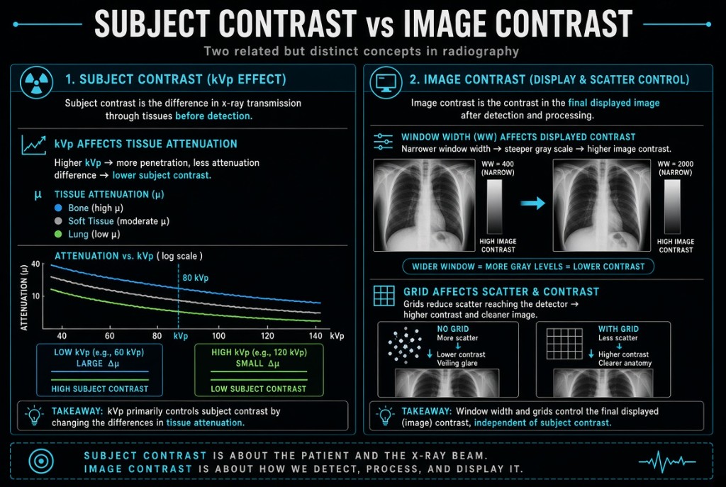
Subject contrast is the difference in x-ray intensity leaving different parts of the patient. It depends on the tissues themselves, meaning their thickness, physical density, and atomic number, and on beam quality. Bone beside muscle, or air-filled lung beside soft tissue, creates high subject contrast. Iodine and barium raise subject contrast because of their high atomic numbers.
Subject Contrast vs Image Contrast
Why it happens
kVp is the main technical factor that controls subject contrast. At lower kVp, the photoelectric effect is more common, and it depends strongly on atomic number, so tissues absorb very differently. At higher kVp, Compton scattering dominates. It depends little on atomic number, so absorption differences shrink and subject contrast drops. Higher kVp also sends more scatter toward the receptor, adding an even gray to the image.
Image contrast
Image contrast is the difference in brightness between areas of the displayed image. It begins with subject contrast, then is shaped by everything after the patient. Scatter reaching the receptor lowers it, so grids and tight collimation raise it. In digital radiography, the look-up table applied during processing largely sets displayed contrast, and window width can change it further at the workstation.
In practice
Because processing rescales the data, kVp changes in DR alter displayed contrast less than they did with film. That does not make kVp unimportant. A beam too weak to penetrate the part cannot be fixed by processing, and excess scatter still degrades the image. Choose kVp for penetration and appropriate subject contrast, control scatter, then let processing and display finish the job.
Clinical Example
A PA chest done at 120 kVp has low subject contrast, which lets lung markings, ribs, and the mediastinum be seen at once. A hand done at 60 kVp shows high subject contrast between cortex and soft tissue. Each is processed with an exam-specific look-up table that sets its displayed contrast.
Common Mistakes
- Saying collimation raises subject contrast, when it reduces scatter and so raises image contrast.
- Assuming window width changes the patient's subject contrast; it changes only the displayed image.
- Believing kVp no longer matters in DR because processing adjusts contrast.
- Forgetting that contrast media work by increasing subject contrast.
Why It Matters
Knowing where contrast comes from tells you which fix to use: kVp and contrast media for subject contrast, scatter control and processing for image contrast. The registry tests which factors belong to each and how kVp affects them.
Key Relationships
- Lower kVp → more photoelectric absorption → higher subject contrast
- Higher kVp → more Compton scatter → lower subject contrast
- Grid or tighter collimation → less scatter → higher image contrast
- Narrower window width → higher displayed contrast