
Image Production · Geometry and detail2 min read5-question check
Spatial resolution
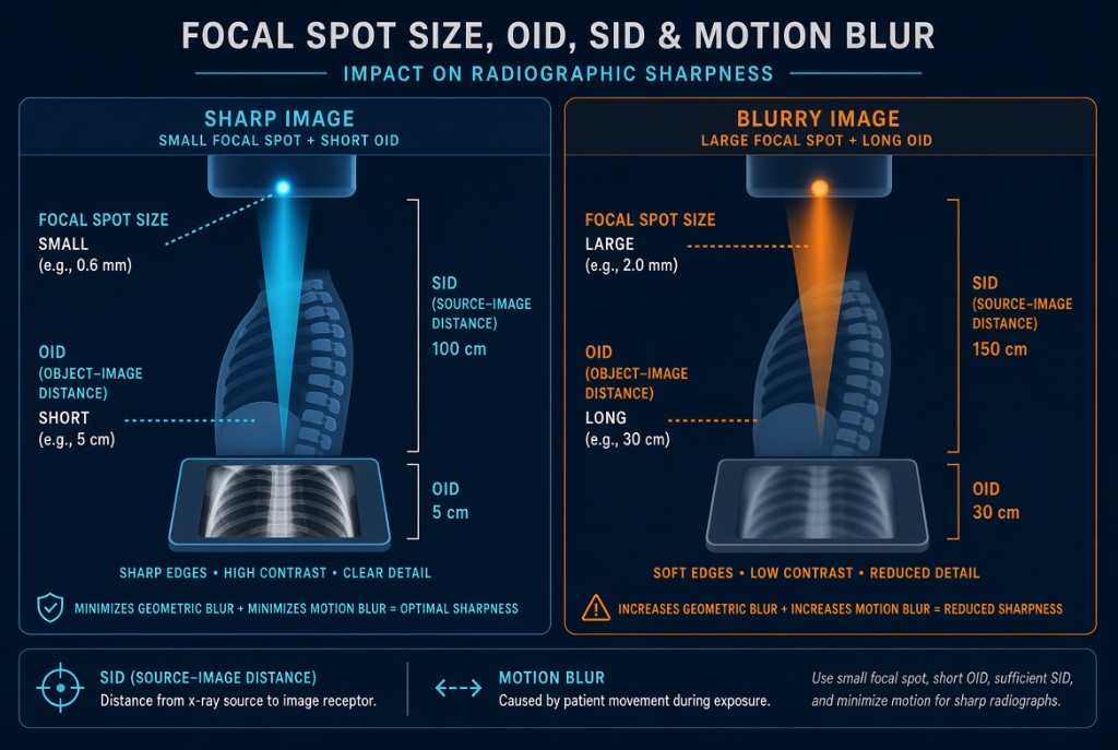
Small focal spot, long SID, short OID, no motion, and small detector elements give sharp detail.
The rule
Spatial resolution is the ability to show small, closely spaced structures as separate, with sharp edges. Film texts called it recorded detail. Five things control it: focal spot size, SID, OID, motion, and the size of the detector elements. kVp and mAs do not directly control it.
Factors Affecting Spatial Resolution
Why it happens
X-rays come from a focal spot with real width, not a single point. Each edge in the anatomy is projected from slightly different points on the target, so it is recorded with a blurred fringe called penumbra, or geometric unsharpness. A smaller focal spot narrows that fringe. A longer SID makes the rays more nearly parallel. A shorter OID gives the rays less distance to spread after they pass the part. Motion often outweighs all of these: voluntary motion, breathing, and involuntary motion such as heartbeat and peristalsis. Fix it with clear instructions, immobilization, and the shortest practical exposure time.
The numbers
Geometric unsharpness equals focal spot size times OID, divided by SOD, the source-to-object distance. Worked example: a 1.2 mm focal spot, 40 inch SID, and 4 inch OID give an SOD of 36 inches. Unsharpness is 1.2 times 4 divided by 36, about 0.13 mm. Switch to a 0.6 mm small focal spot and it halves to about 0.07 mm. Keep the large focal spot but cut OID to 2 inches and it falls to about 0.06 mm.
The detector
In digital radiography, the detector sets an upper limit. Each detector element, or DEL, records one pixel value, so smaller DELs and a smaller pixel pitch can resolve finer detail, measured in line pairs per millimeter. A fine detector cannot recover blur already created by geometry or motion. The small focal spot also has a lower mA limit, so on a thick part the longer exposure time can cost more in motion than the small focal spot gains.
Clinical Example
A PA hand is ordered to look for a subtle fracture. You use the small focal spot, place the hand flat on the receptor at 40 inches, and ask the patient to hold still. Each choice shrinks penumbra or motion blur.
Common Mistakes
- Believing more mAs or kVp makes the image sharper.
- Forgetting that a longer SID improves sharpness as well as reducing magnification.
- Expecting a small-pixel detector to fix motion blur.
- Choosing the small focal spot for a thick part where the longer time invites motion.
Why It Matters
Fine fractures and small lesions are missed when edges blur. The registry asks which factor change improves or degrades spatial resolution and how geometric unsharpness is calculated.
Key Relationships
- Smaller focal spot → sharper image
- Longer SID or shorter OID → sharper image
- Unsharpness = focal spot × OID ÷ SOD
- Smaller DEL → higher maximum resolution
- Motion → blur that nothing else can fix