
Procedures · Chest, abdomen and GI3 min read5-question check
Soft tissue neck, airway and swallowing studies
Thumb sign on the lateral is epiglottitis, steeple sign on the AP is croup; aspiration means barium went below the vocal cords.
Soft tissue neck
The soft tissue neck (upper airway) series is an AP and a lateral, taken erect when possible with the air-filled airway as natural contrast. The lateral is the key view: a true lateral with the shoulders pulled down, chin slightly raised, at 72 inches SID to reduce magnification. Center to about C6 and C7, midway between the laryngeal prominence and the jugular notch, and include from the external acoustic meatus down to the upper thorax. Expose during a slow, deep inspiration so the trachea is filled with air, and use a soft tissue technique, lower than for a C-spine, so the epiglottis and prevertebral soft tissue are not burned out.
Lateral soft tissue neck: normal anatomy and the thumb sign
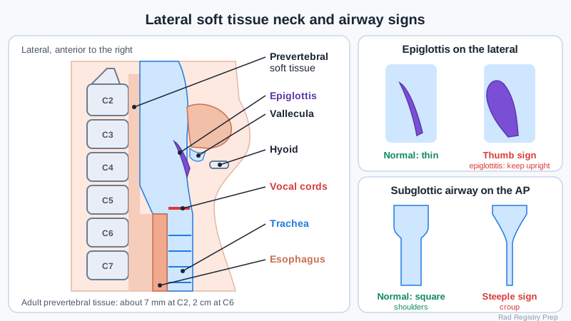
Swollen airway
Three findings are classic. A swollen, rounded epiglottis on the lateral is the thumb sign of epiglottitis, a medical emergency that can close the airway. Keep the patient sitting upright, never lay them flat, take only a lateral, and have the airway team with the patient. Croup narrows the airway just below the vocal cords, which looks like a church spire on the AP: the steeple sign. A retropharyngeal abscess widens the prevertebral soft tissue in front of the upper C-spine; in adults it should be no more than about 7 mm at C2 and about 2 cm at C6.
Foreign bodies
For a swallowed foreign body in a child, AP and lateral films from the nasopharynx to the anus (a foreign body survey) find radiopaque objects. A coin in the esophagus lies flat in the coronal plane, so it looks round on the AP and like a thin line on the lateral. A coin in the trachea usually does the opposite. Button batteries show a double-ring halo and are an emergency. An aspirated object that is not radiopaque, such as a peanut, is found by air trapping: take PA chests on inspiration and expiration, or bilateral decubitus views in small children. The lung with the object stays dark on expiration and the mediastinum shifts away from it.
Swallowing study
The swallowing dysfunction study, also called a modified barium swallow (MBS) or videofluoroscopic swallow study, is done with a speech-language pathologist. The patient sits upright in a lateral position first, then AP, while swallowing barium mixed into thin liquid, thickened liquid, purée and a solid such as a cracker. Record with continuous or high frame rate fluoroscopy from the lips to the cervical esophagus (about C7). Penetration means barium enters the laryngeal vestibule but stays above the vocal cords. Aspiration means it passes below the cords into the trachea. A standard esophagram is a different exam, done RAO to look at the esophagus itself.
Clinical Example
A drooling, feverish child is brought in sitting upright and leaning forward. You take a single erect lateral soft tissue neck without laying the child down, with the ED physician present. The film shows a thumb-shaped epiglottis, and the child goes straight to the OR for airway control.
Common Mistakes
- Laying a child with suspected epiglottitis supine for an AP; it can close the airway.
- Using a C-spine technique for a soft tissue neck, which burns out the epiglottis and airway.
- Expecting a peanut to show on a routine chest; non-radiopaque objects need inspiration and expiration views.
- Mixing up penetration and aspiration on a swallowing study; aspiration is below the vocal cords.
Why It Matters
The 2027 outline lists soft tissue neck, airway and ingested foreign bodies and the swallowing dysfunction study. These exams change patient care fast, and the registry tests the signs, the safe positioning and the projections.
Key Relationships
- Thumb sign (lateral) → epiglottitis; keep the patient upright
- Steeple sign (AP) → croup (subglottic narrowing)
- Widened prevertebral soft tissue → retropharyngeal abscess
- Coin round on AP, edge-on on lateral → esophagus
- Radiolucent aspirated object → inspiration and expiration (air trapping)
- MBS: penetration above cords, aspiration below cords