
Procedures · Head, spine and pelvis3 min read5-question check
Skeletal (osseous) survey
Every region gets its own collimated, marked image: about 20 views for a child, a planned set for myeloma, and never one babygram.
Why and when
A skeletal survey, also called an osseous or bone survey, is a planned set of radiographs that covers most of the skeleton. The three main reasons are suspected non-accidental trauma (NAT) in a young child, a cancer that destroys bone such as multiple myeloma, and suspected skeletal dysplasia (an inherited bone growth disorder). Each reason has its own list of views, but the rules are the same: every region is a separate, tightly collimated image with a lead marker, taken with enough detail to show a tiny fracture line.
Child abuse skeletal survey: separate collimated views
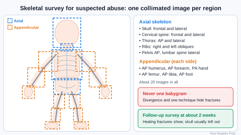
The child abuse survey
For suspected abuse the survey is routine in children under 2 and selective from 2 to 5. Following the ACR protocol, the axial skeleton is a frontal and lateral skull, frontal and lateral cervical spine, AP and lateral thorax, right and left oblique ribs, AP pelvis and lateral lumbar spine. The appendicular skeleton is an AP of each humerus, forearm, femur and tibia, a PA of each hand and an AP of each foot. That is about 20 images. Fractures that raise concern are posterior rib fractures (from squeezing the chest), classic metaphyseal lesions at the ends of long bones (corner or bucket-handle fractures), scapula, sternum and spinous process fractures, and several fractures of different ages. A follow-up survey about 2 weeks later finds fractures that only show once they start to heal. The skull is usually left out of it, because skull fractures do not form callus.
Adult surveys
In adults, a myeloma survey covers the bones that hold red marrow. A typical set is a PA chest, lateral skull, AP and lateral cervical, thoracic and lumbar spine, AP pelvis, and AP humeri and femurs. Myeloma lesions are lytic (punched-out holes), with little new bone forming, so a nuclear medicine bone scan can miss them. Many centers now use whole-body low-dose CT instead, but the radiographic survey is still on the exam. A metastatic survey is less common now, because a bone scan finds blastic metastases from prostate or breast cancer more easily. A dysplasia survey adds views such as a PA left hand for bone age and a lateral thoracolumbar spine.
Doing it well
A whole-body babygram is never acceptable. Beam divergence distorts the edges of the field, and one exposure cannot be right for both the skull and the feet. Use a small focal spot, a technique set for each part, and the smallest field that covers it. Check every image for rotation and clipped joints before the patient leaves, since a repeat days later can miss findings. Myeloma patients have fragile bones, so move them slowly and let them help. With a child, stay neutral and kind with the family, record who was present and anything you saw, and follow facility policy and state law for reporting. Never discuss suspected abuse with the caregivers yourself.
Clinical Example
A 4-month-old comes from the ED with a swollen thigh and a story that does not fit. You collimate each region, taking both rib obliques and an AP of each hand and foot, and check every image before the family leaves. The radiologist finds healing posterior rib fractures that no one suspected. A follow-up survey is booked for 2 weeks.
Common Mistakes
- Taking one or two whole-body images instead of separate, collimated views.
- Leaving out the rib obliques, which are the best views for posterior rib fractures.
- Thinking a normal bone scan rules out multiple myeloma.
- Talking with caregivers about suspected abuse instead of documenting and following policy.
Why It Matters
The 2027 ARRT outline lists the bone survey as a projection the registry can ask about. Questions ask who gets one, which views it includes, why it is not a babygram, and why it is used for myeloma.
Key Relationships
- Suspected NAT, under 2 → full skeletal survey (about 20 images)
- Posterior rib fractures + metaphyseal corner fractures → strong concern for abuse
- Follow-up survey ~2 weeks → healing fractures show
- Myeloma = lytic → bone scan may miss it → survey or low-dose CT
- Babygram → divergence and one technique for all → not acceptable