
Image Production · Geometry and detail2 min read5-question check
Size Distortion vs Shape Distortion
Size distortion enlarges the whole part evenly; shape distortion stretches or shrinks one dimension.
Two kinds
Distortion is any difference between the real size or shape of a part and how it appears on the image. Size distortion is magnification. It comes from SID and OID, and it makes the whole part larger in every direction by the same amount. Shape distortion is unequal magnification of different parts of the same object. It shows up as elongation, where the part looks longer than it is, or foreshortening, where it looks shorter.
Size vs Shape Distortion
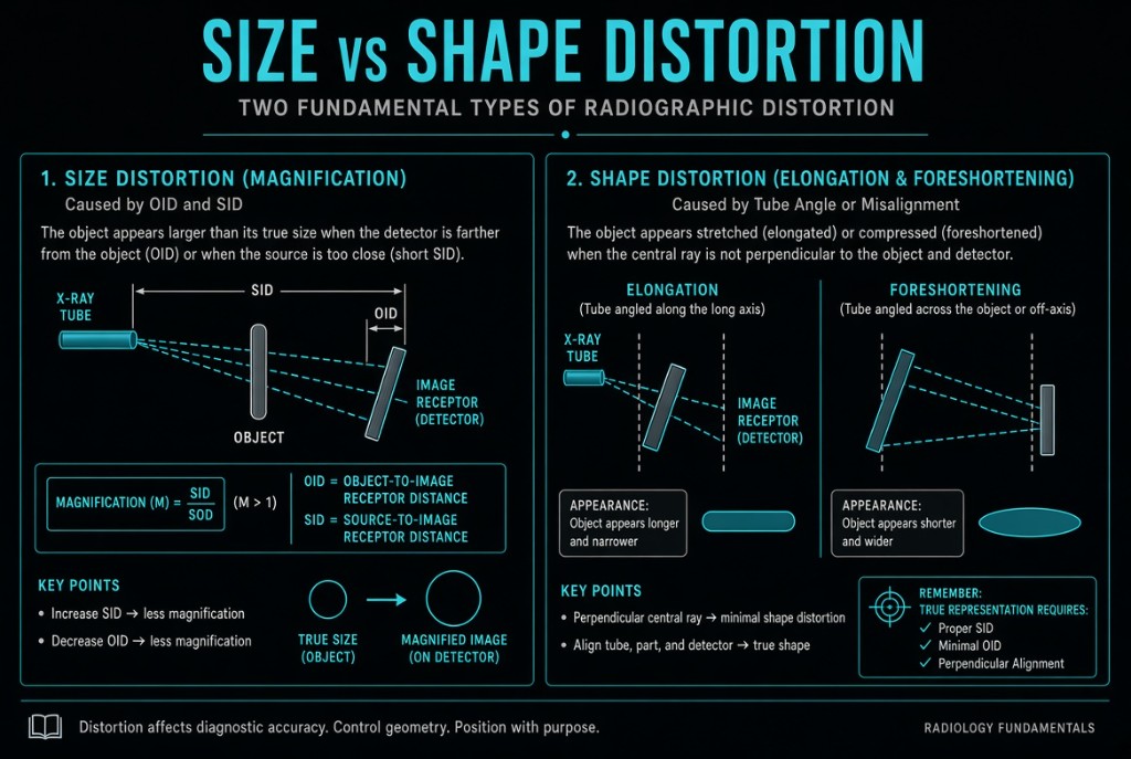
How shape distortion happens
Shape distortion comes from misalignment between the tube, the part, and the image receptor. Angling the tube or the receptor causes elongation. Tilting the part so it is no longer parallel to the receptor causes foreshortening. Centering also matters. Anatomy near the central ray is projected by fairly straight rays, while anatomy far from the center is struck by divergent rays and can be distorted, so center the central ray to the area of interest.
Intentional angles
Not all shape distortion is a mistake. Radiographers angle the central ray on purpose to open up anatomy or to move overlapping structures apart. The PA axial scaphoid projection angles toward the elbow to elongate the scaphoid so a fracture line is easier to see. The AP axial projection of the skull angles caudad to project the occipital bone clear of the facial bones. In these cases the elongation is the goal.
Telling them apart
To tell the two apart, look at proportions. If a structure is uniformly bigger but has its normal shape, the cause is size distortion, so check SID and OID. If one dimension is stretched or squashed while the other looks normal, the cause is shape distortion, so check the tube angle, part alignment, and centering. Changing SID alone never creates elongation or foreshortening.
Clinical Example
On a lateral knee, the femoral condyles fail to superimpose and the knee looks unnaturally short top to bottom. The leg was tilted rather than parallel to the detector. Repositioning the part parallel to the receptor fixes the foreshortening without changing distance.
Common Mistakes
- Trying to fix elongation by increasing SID, which only reduces magnification.
- Assuming every angled projection is a positioning error when many angles are planned.
- Mixing up the causes: an angled tube elongates, while a tilted part foreshortens.
- Centering away from the area of interest and ignoring the distortion from divergent rays.
Why It Matters
Distorted anatomy can hide fractures, mimic disease, or ruin measurements. The registry often shows a scenario and asks which factor caused the change in appearance.
Key Relationships
- Size distortion = magnification, controlled by SID and OID
- Shape distortion = elongation or foreshortening from misalignment
- Angled tube or receptor → elongation
- Part not parallel to receptor → foreshortening
- Farther from the central ray → more shape distortion