
Image Production · Geometry and detail2 min read5-question check
Size distortion and magnification
Magnification grows with OID and shrinks with SID; magnification factor is SID divided by SOD.
The rule
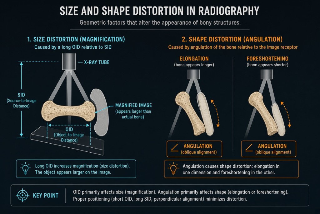
Distortion is any misrepresentation of the true size or shape of anatomy. Size distortion is magnification: the image is larger than the object. Every radiograph has some, because the part always sits some distance above the receptor and x-rays diverge from the focal spot. Two distances control it, SID and OID. Shape distortion, meaning elongation and foreshortening, comes from alignment and angulation and is a separate topic.
Size and Shape Distortion
Why it happens
Diverging rays keep spreading after they pass through the part. The farther the part is from the receptor, the more they spread before the image is recorded, so increasing OID increases magnification. A longer SID makes the rays passing the part more nearly parallel, so increasing SID decreases magnification. Magnification costs spatial resolution too, because the same geometry widens penumbra. When the part is parallel to the receptor and the central ray is aligned, magnification changes size but not shape.
The numbers
Magnification factor equals SID divided by SOD, where SOD is SID minus OID. Worked example: a 40 inch, or 102 cm, SID with the part 4 inches from the receptor gives an SOD of 36 inches. The factor is 40 divided by 36, about 1.11, so the image is 11 percent larger than the part. A structure measuring 5.5 cm on the image is really 5.5 divided by 1.11, about 5 cm. Raise SID to 72 inches with the same OID and the factor falls to 72 divided by 68, about 1.06.
On the job
This is why a routine chest is done PA at 72 inches (183 cm). The heart lies anterior, so the PA position puts it close to the receptor, and the long SID further limits its magnification. An AP chest makes the heart look larger. Keep the part in contact with the receptor when you can. When OID cannot be avoided, as with a cross-table lateral or a patient who cannot flatten a joint, increase SID to compensate.
Clinical Example
A portable AP chest at 40 inches shows a heart that looks enlarged. Because the heart is far from the receptor and the SID is short, much of that size is magnification. The radiologist reads cardiac size with that in mind, and the next upright exam is done PA at 72 inches.
Common Mistakes
- Thinking a longer SID increases magnification.
- Dividing by SID instead of SOD when finding object size.
- Calling magnification shape distortion.
- Accepting extra OID without raising SID to compensate.
Why It Matters
Magnification distorts measurements such as heart size and lowers sharpness. The registry tests the SID and OID relationships and asks you to calculate magnification factor or true object size.
Key Relationships
- Increase OID → more magnification
- Increase SID → less magnification
- MF = SID ÷ SOD
- Object size = image size ÷ MF