
Image Production · Geometry and detail2 min read5-question check
SID versus OID
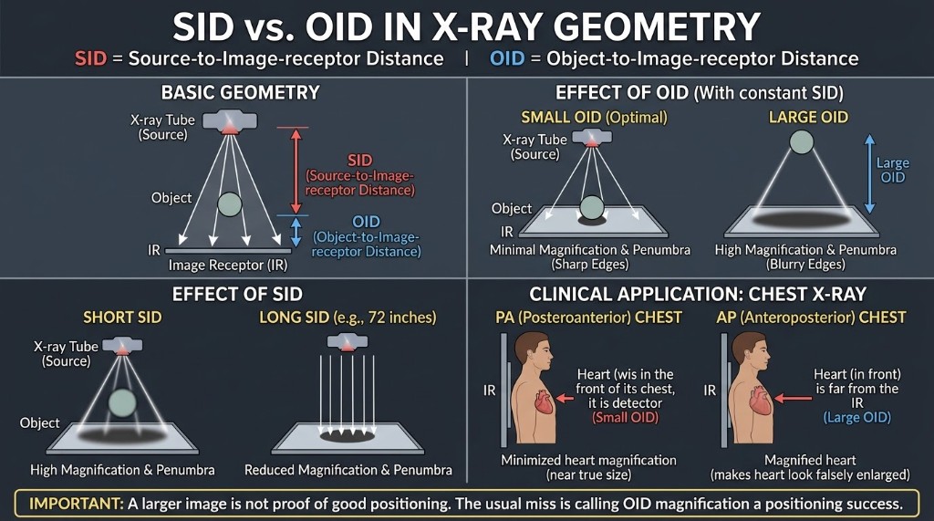
Long SID and short OID mean less magnification and sharper edges.
The terms
SID is source-to-image receptor distance, measured from the focal spot to the receptor. OID is object-to-image receptor distance, measured from the part to the receptor. A third distance, SOD, is source-to-object distance, which equals SID minus OID. Together these set how much the image is magnified and how sharp its edges are.
How it works
X-rays diverge from the focal spot, so any part held away from the receptor projects a larger shadow. More OID means more magnification. A longer SID makes the rays striking the part closer to parallel, which reduces magnification. Because the focal spot has real size, OID also widens the penumbra, the blurred edge around each structure, and lowers spatial resolution. Keep the part close to the receptor and use the longest practical SID. A longer SID also lowers receptor exposure by the inverse square law, so it needs more mAs.
The numbers
Magnification factor equals SID divided by SOD. Worked example: at 40 inches SID, a bone 4 inches from the receptor has an SOD of 36 inches. 40 divided by 36 is about 1.11, so the image is 11% larger than the bone. At 72 inches with the same 4-inch OID, the factor is 72 divided by 68, about 1.06. To find true size, divide image size by the magnification factor. A 10 cm image at a factor of 1.25 means the object is 8 cm.
On the job
Chest radiography uses 72 inches, about 183 cm, and a PA projection so the heart, an anterior structure, sits close to the receptor. An AP chest places the heart farther from the receptor, so it looks larger, and a mobile AP chest at a shorter SID magnifies it further. A large OID has one benefit: the air gap lets some scatter miss the receptor. The air-gap technique uses this, trading magnification for less scatter without a grid.
Clinical Example
A patient's wrist is propped on a sponge 3 inches above the receptor for a lateral view. Before exposing, you notice the gap and lower the wrist directly onto the receptor. The image will be sharper and closer to true size.
Common Mistakes
- Mixing up the terms: SID starts at the focal spot, OID starts at the part.
- Reading a large heart on an AP chest as true enlargement without considering OID.
- Lengthening SID without raising mAs, which lowers receptor exposure.
- Forgetting to subtract OID from SID before computing the magnification factor.
Why It Matters
Magnification can mimic disease, such as an enlarged heart, and soft edges hide fine detail. The registry asks you to rank setups by magnification and to compute the magnification factor or true object size.
Key Relationships
- SOD = SID − OID
- Magnification factor = SID / SOD
- More OID → more magnification, less spatial resolution
- Longer SID → less magnification, more mAs needed
- Object size = image size / magnification factor