
Procedures · Fluoro and special studies3 min read5-question check
Shunt series (VP shunt)
Follow the tube from the skull to its tip with overlapping images: skull, neck, chest and abdomen, and no gaps.
What a shunt is
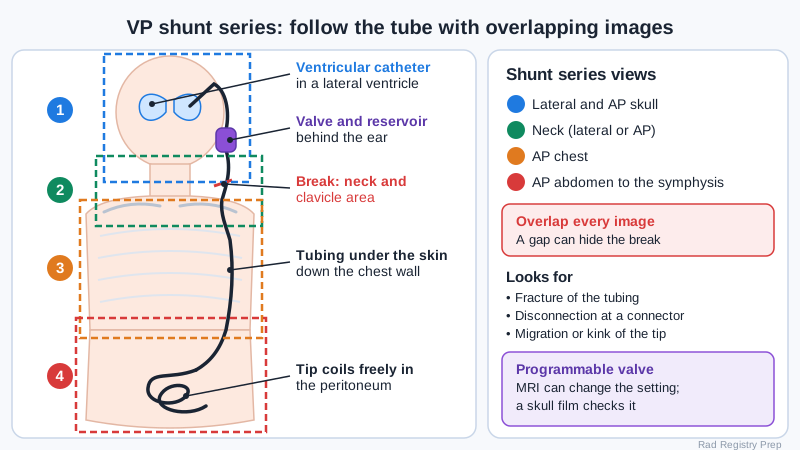
A CSF shunt drains extra cerebrospinal fluid from hydrocephalus. The most common type is the ventriculoperitoneal (VP) shunt. A catheter enters a lateral ventricle through a burr hole, runs to a valve and reservoir under the scalp, usually behind the ear, then tunnels under the skin down the neck, over the clavicle and chest wall, and into the peritoneal cavity, where the fluid is absorbed. A ventriculoatrial (VA) shunt ends in the right atrium instead, and a ventriculopleural shunt in the pleural space. The tubing is radiopaque so radiographs can trace it.
VP shunt path and the overlapping shunt series views
The views
A shunt series is a set of radiographs that follows the whole tube. A common set is a lateral skull and a frontal (AP) skull to show the ventricular catheter and valve, a lateral or AP neck, an AP chest and an AP abdomen, often with the patient supine. Protocols vary, so follow yours, but the rule is the same everywhere: each image must overlap the next so no length of tubing falls in a gap. Turn the head so the valve side is not hidden, and include the whole abdomen down to the pubic symphysis, since the tip can lie low in the pelvis. For a VA shunt, the chest image must show the tip near the right atrium.
What the films show
The series looks for mechanical failure. A fracture (break) in the tubing happens most often where it moves with the neck or crosses the clavicle and lower ribs. A disconnection happens at a connector. Migration means the tip has moved, for example out of the peritoneum, into the chest or scrotum, or up into the subcutaneous tissue. A kink or a short peritoneal catheter in a growing child are other causes. The patient may have headache, vomiting, sleepiness or, in a baby, a bulging fontanelle and a growing head. Radiographs show the hardware. Ventricle size is checked with CT, fast MRI or ultrasound through an open fontanelle.
Doing it well
Many valves are programmable. Their pressure setting is shown by a small magnet and a mark that you can read on a skull radiograph. A strong magnet such as an MRI scanner can change the setting, so a skull film to check the valve is often ordered after MRI. Do not press on the reservoir or the tubing. Lay the patient's head on the side without the valve when you can. Many shunt patients are children who will have this series many times over their lives, so collimate to the tubing, shield when it does not cover the tube, and avoid repeats by checking overlap before the patient leaves.
Clinical Example
A 6-year-old with a VP shunt comes in vomiting and sleepy. You take lateral and AP skull, AP neck, AP chest and AP abdomen, checking that each image overlaps the next. On the neck view the tubing has a clean break just above the clavicle, and neurosurgery takes her to the OR to revise it.
Common Mistakes
- Leaving a gap between images, so the break falls in the length of tubing no one imaged.
- Cutting off the lower abdomen and pelvis where the peritoneal tip lies.
- Pressing on the reservoir or tubing while positioning.
- Forgetting that MRI can change a programmable valve setting, which a skull film then checks.
Why It Matters
Shunt series are routine in pediatric and neuro units, and the registry can ask what the series includes, what it looks for, and how to care for the patient. A missed gap can mean a missed break and a sick child.
Key Relationships
- VP shunt: lateral ventricle → valve behind ear → neck → chest wall → peritoneum
- Shunt series: skull (lat + AP) → neck → chest → abdomen, all overlapping
- Tubing break → most often neck or clavicle area
- Disconnection → at connectors
- Programmable valve + MRI → skull film to check the setting
- VA shunt tip → right atrium