
Procedures · Extremities3 min read5-question check
Shoulder film check
External rotation puts the greater tubercle in profile, internal rotation the lesser, Grashey opens the joint, and the Y or axillary shows where the head really sits.
External and internal rotation
For the AP shoulder in external rotation, turn the hand palm up so the epicondyles are parallel to the IR. The greater tubercle shows in profile on the lateral edge of the humeral head. For internal rotation, turn the hand palm down, back of the hand on the hip, so the epicondyles are perpendicular. The lesser tubercle shows in profile on the medial side, and the humeral head looks rounder. Center about 1 inch below the coracoid. In trauma, do not rotate the arm: take a neutral AP and a second view instead.
Grashey
The Grashey, an AP oblique, turns the patient 35 to 45 degrees toward the affected side so the scapula is parallel to the IR. On a good Grashey, the glenoid cavity is in profile and the glenohumeral joint space is open, with no overlap of the humeral head and the glenoid. If the humeral head still overlaps the glenoid, the rotation was off for this patient. Every patient's chest is shaped a little differently, so adjust the oblique and check the joint space again.
Scapular Y
The scapular Y is a PA oblique with the patient turned 45 to 60 degrees, affected side to the IR. The scapula is seen on end as a Y: the body forms the stem, the acromion and coracoid the arms. When the shoulder is in joint, the humeral head sits over the center of the Y. An anterior dislocation puts the head below the coracoid; a posterior dislocation puts it below the acromion. If the scapular body is not a thin line, adjust the rotation.
Axillary and trauma
A posterior dislocation can look almost normal on an AP. That is why every shoulder series needs a second view at a right angle, a Y or an axillary. The inferosuperior axillary shows the humeral head and the glenoid from below, so you can see whether the head is in front of or behind the glenoid. Abduct the arm only as far as the patient can tolerate, and never force it in trauma.
Read the films
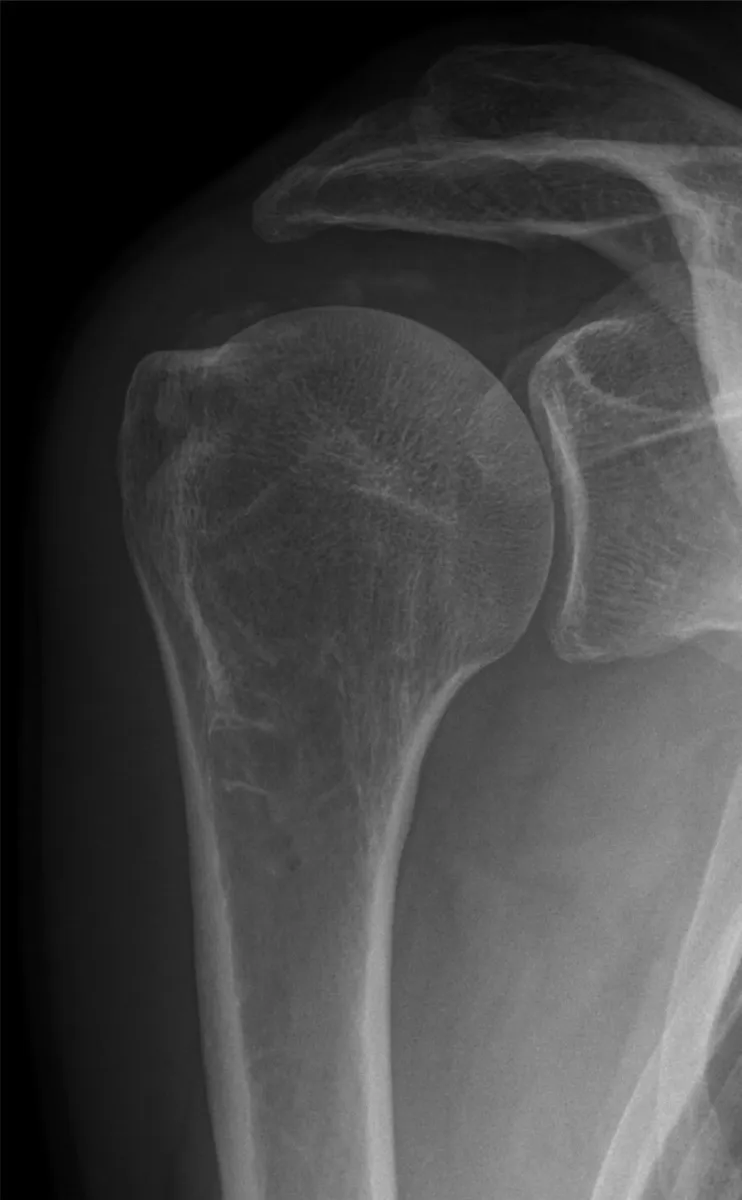
KeeperAP shoulder, external rotation
Look for: The greater tubercle is in profile on the lateral side of the humeral head. This patient also has calcium in the supraspinatus tendon, which this view shows well.
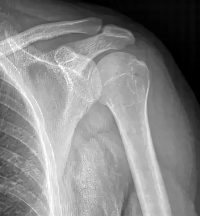
KeeperAP shoulder, internal rotation
Look for: The humeral head looks rounder and the greater tubercle no longer shows in profile laterally. The lesser tubercle turns toward the medial side.
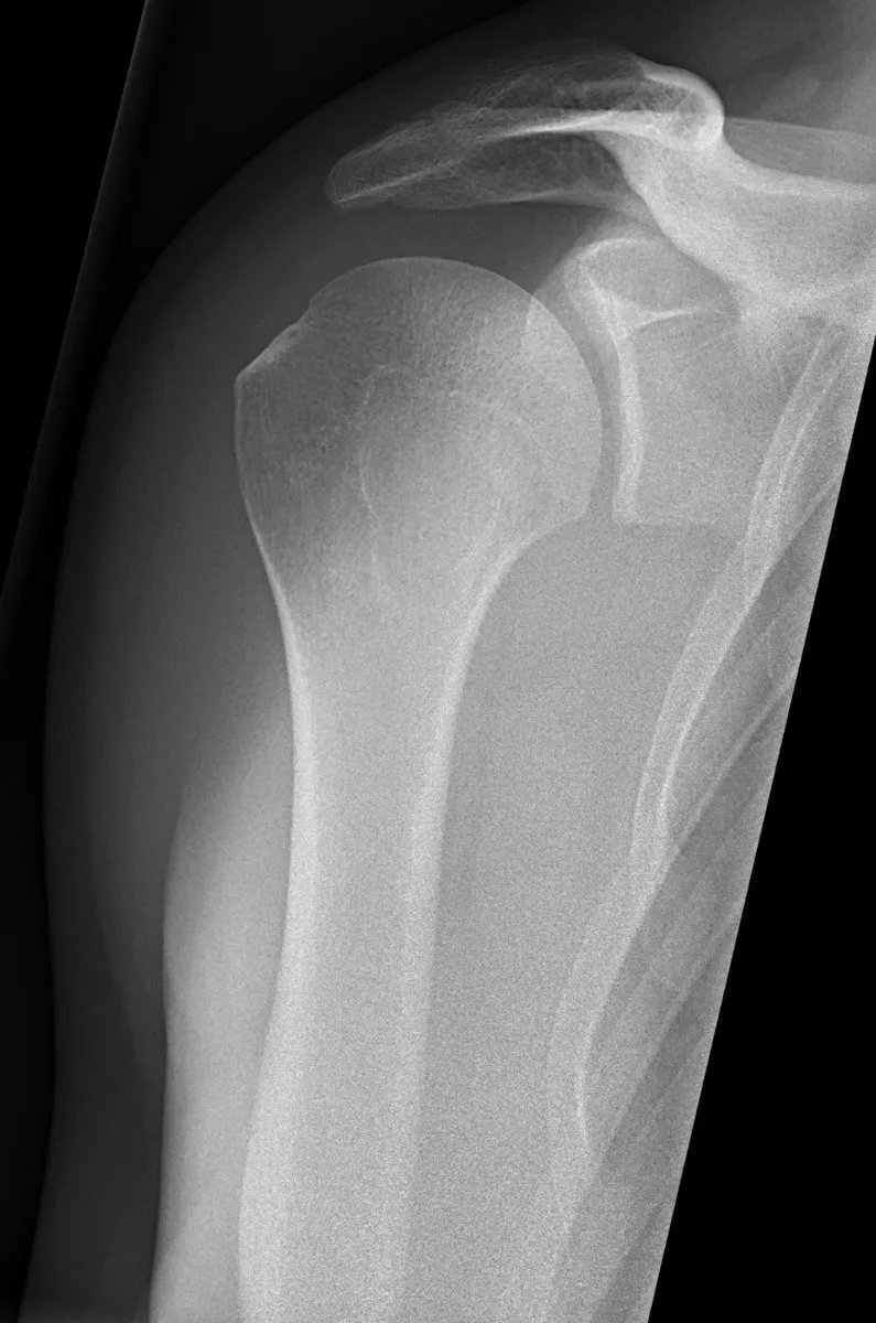
KeeperGrashey: a keeper
Look for: The glenoid is in profile and the glenohumeral joint space is open, with no overlap of the humeral head on the glenoid.
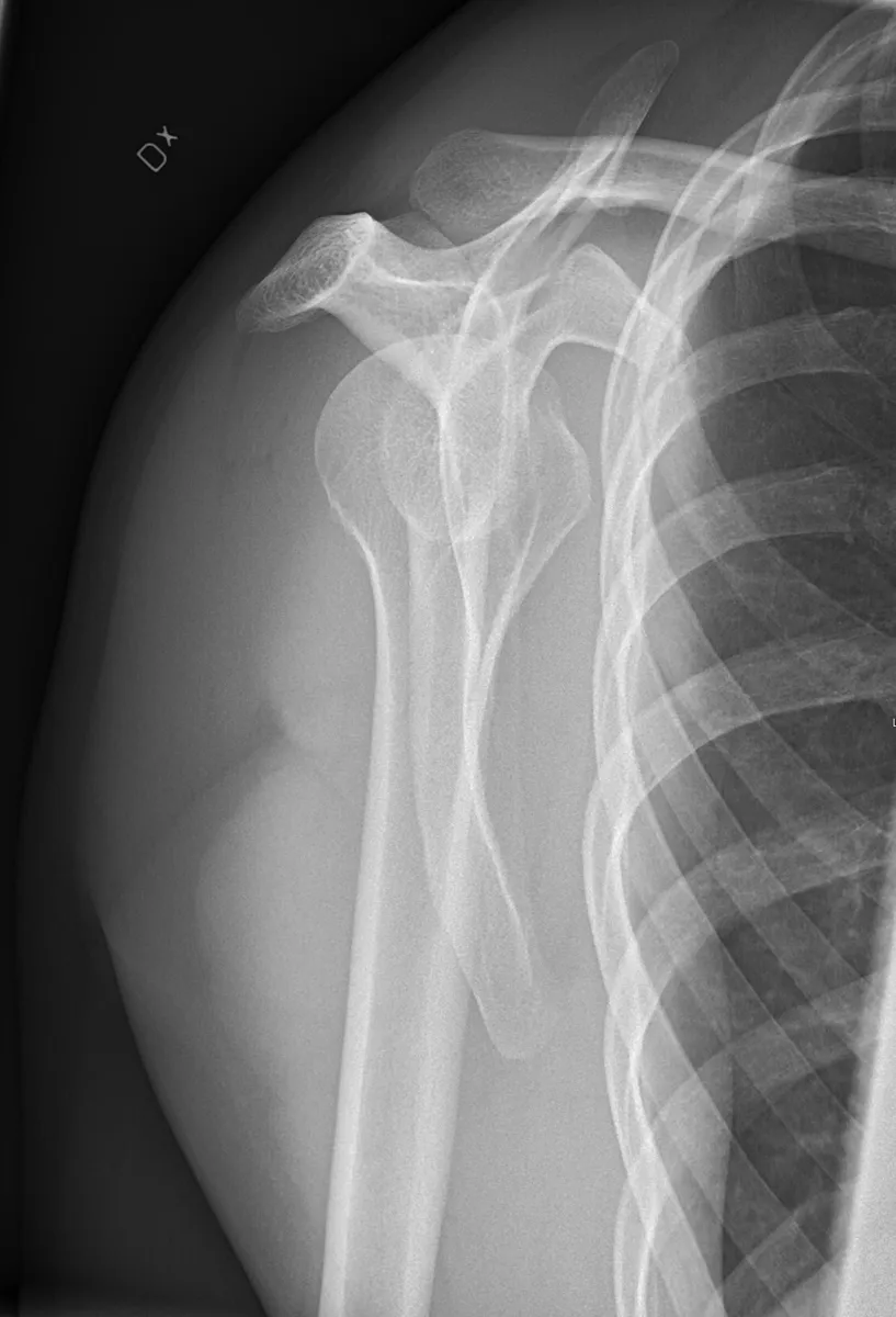
KeeperScapular Y: a keeper
Look for: The scapular body is on end as the stem of the Y, and the humeral head sits over the center of the Y. The shoulder is in joint.
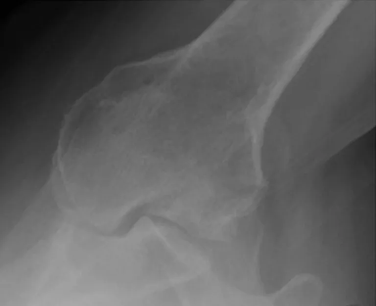
KeeperAxillary: why the second view matters
Look for: On the axillary, the humeral head sits behind the glenoid: a posterior dislocation. It can look almost normal on the AP, so the second view made the diagnosis.
Clinical Example
A seizure patient holds the arm in internal rotation and the AP looks almost normal. Because seizures are a classic cause of posterior dislocation, you do not rotate the arm. You take a scapular Y and an axillary as far as the patient can abduct, which shows the humeral head behind the glenoid.
Common Mistakes
- Rotating the arm for external and internal views on a trauma patient.
- Accepting a Grashey where the humeral head overlaps the glenoid; adjust the oblique until the joint space opens.
- Calling the shoulder normal from the AP alone.
- Mixing up the dislocation signs on the Y; anterior goes below the coracoid, posterior below the acromion.
Why It Matters
Missed posterior dislocations are a classic error, and the registry tests which view shows what: tubercles on the rotation views, the glenoid on the Grashey, and dislocation on the Y and axillary.
Key Relationships
- External rotation → greater tubercle in profile laterally
- Internal rotation → lesser tubercle in profile medially
- Grashey 35 to 45° → glenoid in profile, open joint
- Scapular Y: head below the coracoid → anterior dislocation
- Scapular Y: head below the acromion → posterior dislocation