
Image Production · Scatter, grids and filtration2 min read5-question check
Scatter and collimation
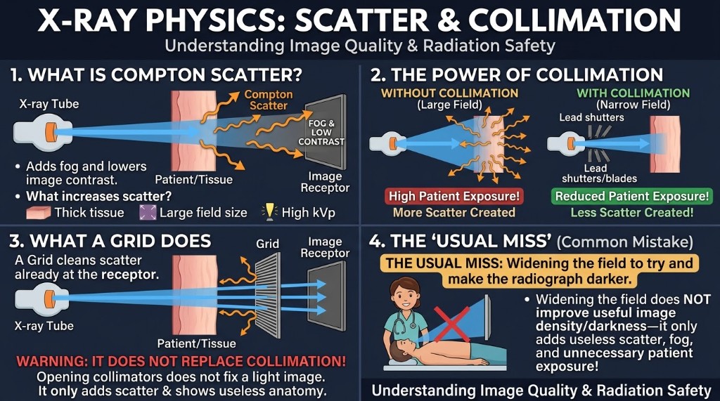
Collimation shrinks the volume of tissue irradiated, cutting both scatter and patient dose.
The rule
Scatter is radiation that changes direction inside the patient, mostly through Compton interactions. It carries no useful information. When it reaches the receptor it adds fog, lowers contrast, and hides low-contrast detail. Three factors increase scatter: higher kVp, larger field size, and greater part thickness. Collimation is your most direct control over how much is produced.
Why it happens
Scatter is created wherever the primary beam strikes tissue. A larger field irradiates more tissue, so more Compton interactions occur and more scatter heads toward the receptor. Restricting the beam to the anatomy of interest shrinks that volume. Less scatter is produced, contrast improves, and the patient receives less dose because less tissue is exposed. A grid works differently. It absorbs scatter after it has left the patient, so it adds to collimation but never replaces it.
How it works
The variable-aperture collimator is the standard beam restrictor. Lead shutters set the field size, and a light and mirror show where the beam will fall. The light field and x-ray field must line up within 2% of the SID. At 40 inches that is 0.8 inch. Cones and cylinders are fixed devices still used for some small fields. Electronic masking or shuttering after the exposure only changes how the image is displayed. It does not reduce scatter or dose and must never replace collimation.
On the job
Collimate to the anatomy of interest, with the field borders visible on the image. Opening the field does not fix a low exposure indicator. It adds scatter and dose. Accurate collimation also helps the system recognize the exposed area, which keeps the exposure indicator reliable. With AEC, tighter collimation may lengthen the exposure slightly because less scatter reaches the chambers, which is expected.
Clinical Example
An AP lumbar spine is set up with the field opened to the full 14 by 17 inch receptor. You narrow the side-to-side field to include the vertebral bodies, transverse processes, and SI joints. Less tissue is irradiated, scatter drops, and the spine shows better contrast at a lower dose.
Common Mistakes
- Opening the field to improve a low exposure indicator, which only adds scatter and dose.
- Relying on electronic masking instead of collimating before the exposure.
- Assuming a grid makes collimation unnecessary.
- Collimating so tightly that required anatomy is cut off, forcing a repeat.
Why It Matters
Collimation improves contrast and lowers patient dose at the same time. The registry asks which factors increase scatter, what collimation does to contrast and dose, and the light field accuracy standard.
Key Relationships
- Larger field, higher kVp, thicker part → more scatter
- Tighter collimation → less scatter, higher contrast, lower dose
- Grid removes scatter leaving the patient; collimation prevents it
- Light field and x-ray field must align within 2% of SID