
Procedures · Head, spine and pelvis3 min read5-question check
Pelvis and hip film check
Legs turned in 15 to 20° so the femoral necks lengthen and the lesser trochanters almost disappear, unless a fracture is possible. Then nothing gets rotated.
The AP pelvis
On the AP pelvis, turn both legs in 15 to 20 degrees. That puts the femoral necks parallel to the IR so they show at full length, and it hides the lesser trochanters behind the femoral shafts, or leaves only a small edge showing. Center midway between the ASIS and the symphysis pubis. The iliac wings and obturator foramina should look the same on both sides. If one obturator foramen looks narrower, the pelvis was rotated. Large lesser trochanters and short femoral necks mean the legs were turned out.
Trauma first
If a hip fracture is possible, do not turn the legs. A fractured hip often lies turned out, with the leg shortened, and rotating it can move the fracture and hurt the patient. Take the AP pelvis as the legs lie, then a cross-table lateral of the hip. The frog-leg lateral, which turns the leg out, is only for patients without trauma.
Inlet and outlet
The inlet and outlet views show the pelvic ring for trauma. The AP axial inlet angles the CR about 40 degrees caudad and looks down into the pelvic ring, showing whether the ring has shifted forward or back. The AP axial outlet angles cephalad, about 20 to 35 degrees for men and 30 to 45 degrees for women, and shows the pubic and ischial bones without foreshortening. Neither view needs the patient to move.
The lateral hip
The axiolateral inferosuperior hip, the cross-table lateral, uses a horizontal beam aimed up through the groin, perpendicular to the femoral neck. Raise and support the other leg out of the way, and place the IR against the side of the hip, parallel to the femoral neck. On a good film, the femoral head, neck and greater trochanter show in profile, and the femoral head sits in the acetabulum.
Read the films
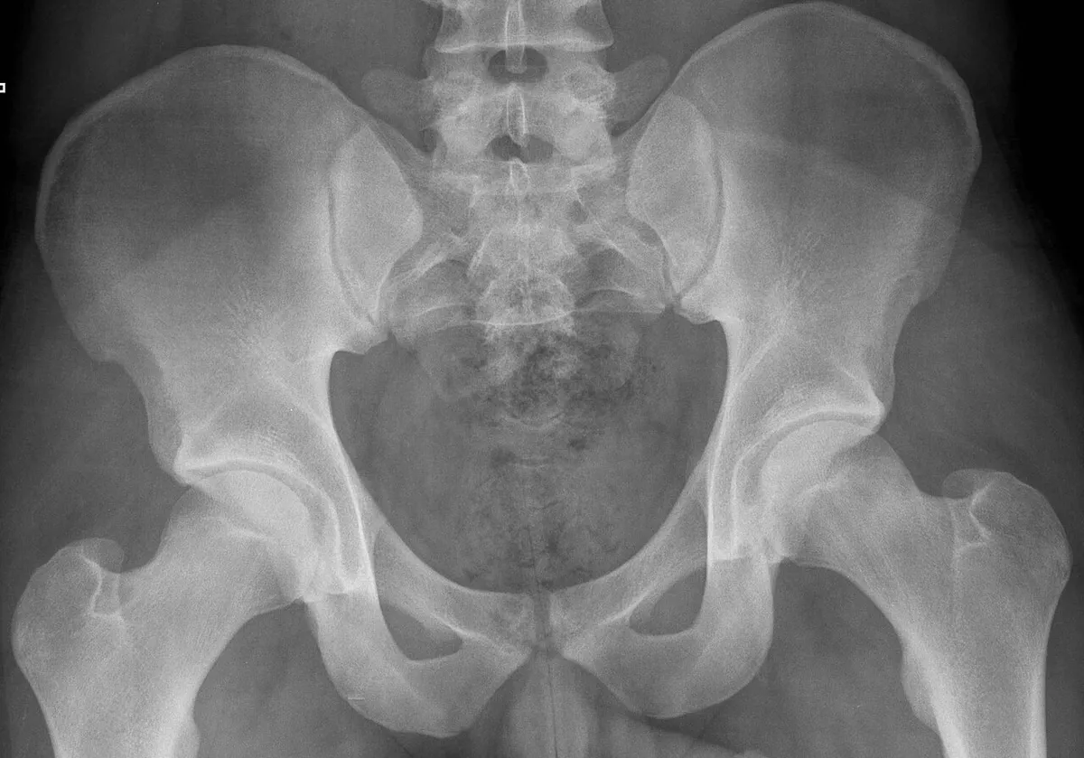
KeeperAP pelvis: a keeper
Look for: The iliac wings and obturator foramina look the same on both sides, and the lesser trochanters are small. The legs were turned in and the pelvis is straight.
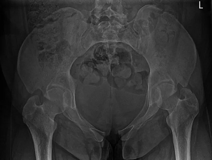
KeeperAP axial inlet: a keeper
Look for: The caudad angle looks down into the pelvic ring, so the ring shows as a round outline. This is the view that shows whether the ring has shifted forward or back.
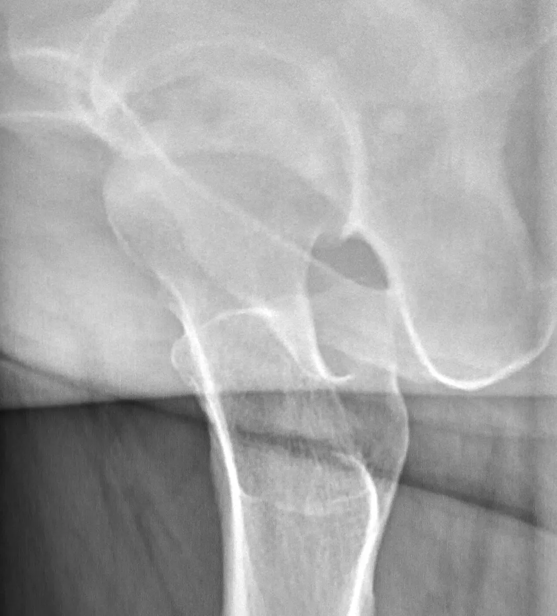
KeeperCross-table lateral hip
Look for: The femoral head and neck are in profile and the head sits in the acetabulum. This patient has avascular necrosis of the femoral head.
Clinical Example
An elderly patient fell at home and the right leg is shortened and turned out. You leave both legs as they lie for the AP pelvis, then take a cross-table lateral of the right hip with the left leg raised on a support. The lateral shows a femoral neck fracture.
Common Mistakes
- Turning the legs in on a patient with a possible hip fracture.
- Doing a frog-leg lateral in trauma.
- Calling large lesser trochanters normal; they mean the legs were turned out.
- Using one angle for the outlet on everyone; women usually need more.
Why It Matters
Hip fractures in elderly patients are one of the most common trauma exams, and moving a fractured hip can displace it. The registry tests the 15 to 20° internal rotation, the inlet and outlet angles, and which lateral to use in trauma.
Key Relationships
- Legs turned in 15 to 20° → femoral necks at full length, lesser trochanters hidden
- Large lesser trochanters → legs turned out
- Possible fracture → do not rotate; cross-table lateral, not frog-leg
- Inlet: about 40° caudad
- Outlet: 20 to 35° cephalad (men), 30 to 45° (women)