
Procedures · Head, spine and pelvis3 min read5-question check
Lumbar spine film check
Spinous processes centered and SI joints even on the AP, open disc spaces on the lateral, and a Scotty dog with its eye in the middle on the obliques.
The AP
For the AP lumbar spine, flex the knees to flatten the lumbar curve and bring the back closer to the IR. Center at the iliac crest, about L4 to L5, for a 14 × 17 that includes T12 to the sacrum. On a straight AP the spinous processes are in the midline, the SI joints are the same distance from the spine, and the pedicles look the same on both sides. If the SI joints are uneven, the pelvis was rotated.
The lateral
The lateral lumbar spine is centered at L4, the level of the iliac crest. Put a support under the waist if needed so the spine is parallel to the table. On a good lateral the intervertebral disc spaces and foramina are open, and the back edges of each vertebral body line up as one line. Doubled back edges mean rotation; stack the shoulders and the hips. Close collimation and a lead blocker behind the patient cut scatter, which matters more on the lateral than almost any other view.
The L5–S1 spot
The L5–S1 spot lateral shows the last joint, which the main lateral often leaves closed. Center about 1.5 inches below the iliac crest and 2 inches behind the ASIS. If the waist is not supported, angle the CR 5 to 8 degrees caudad to line up with the joint. The joint should be open and centered.
The obliques
The obliques show the zygapophyseal joints as the Scotty dog: the pedicle is the eye, the pars interarticularis the neck, and the superior articular process the ear. Turn the patient 45 degrees. Posterior obliques, the RPO and LPO, show the joints on the side closest to the IR. Anterior obliques show the side away from the IR. If the eye sits toward the front of the vertebral body, the patient was not turned enough; toward the back, too much.
Read the films
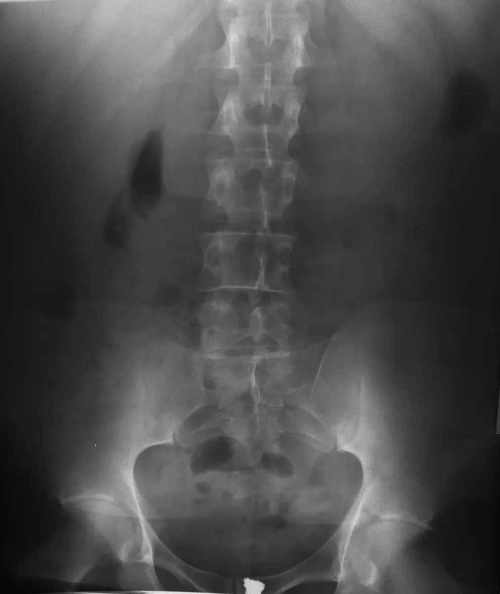
KeeperAP lumbar spine: a keeper
Look for: The spinous processes are in the midline, the SI joints sit the same distance from the spine, and the field runs from the lower thoracic spine to the sacrum.
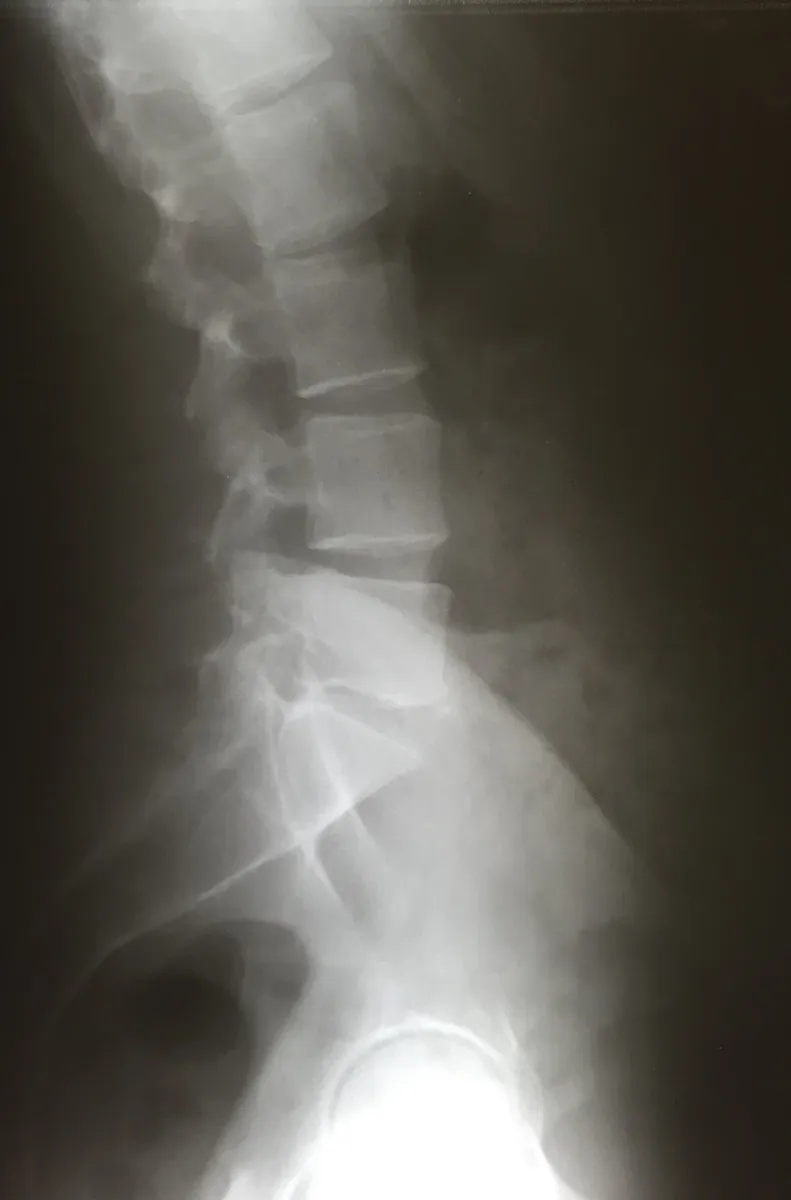
KeeperLateral lumbar spine: a keeper
Look for: The disc spaces are open and the back edges of the vertebral bodies line up, so the patient was not rotated.
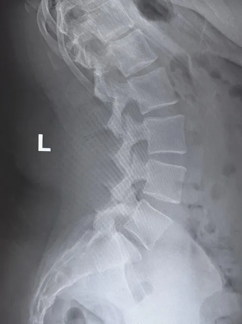
KeeperLateral lumbar, down to L5–S1
Look for: The lower lumbar disc spaces are open down to L5–S1. If the last joint is closed on your lateral, add the L5–S1 spot.
Clinical Example
Your lateral lumbar spine shows the disc spaces open from L1 to L4, but L5–S1 is closed and the back edges of the bodies are doubled. You stack the shoulders and hips, add a support under the waist, and take the L5–S1 spot centered below the crest. Both joints open.
Common Mistakes
- Leaving the knees straight on the AP, which keeps the lumbar curve.
- Calling L5–S1 fine on the main lateral when it is closed; add the spot view.
- Mixing up the obliques: posterior obliques show the side closest to the IR.
- Not using a lead blocker behind the patient on the lateral.
Why It Matters
Lumbar spines are high-dose, scatter-heavy exams, so a repeat costs the patient real dose. The registry tests the Scotty dog parts and which side each oblique shows.
Key Relationships
- AP: knees flexed, CR at the iliac crest
- Uneven SI joints → pelvis rotated
- Lateral: doubled back edges of the bodies → rotation
- L5–S1 spot: 5 to 8° caudad if the waist is not supported
- Posterior oblique → side closest to the IR; Scotty dog eye = pedicle