
Procedures · Extremities4 min read5-question check
Long bones film check: humerus, femur and tib-fib
Both joints on every long bone, two views at right angles, cathode at the thick end, and no rotating an arm or leg that might be broken.
Both joints, two views
Every long bone gets two views at right angles, and on the first exam both views must include the joints at each end. On a follow-up, the joint nearest the injury is often enough. A fracture of one bone can come with a dislocation or a second fracture at the far joint. If the bone is too long for one IR, place it diagonally, or take two overlapping images: one with each joint. Put the cathode end of the tube at the thicker end of the part, the shoulder, hip or knee, to even out the density.
Humerus
For the AP humerus, the epicondyles are parallel to the IR, which puts the greater tubercle in profile. For the lateral, the epicondyles are perpendicular to the IR, with the hand turned in. Both should include the shoulder and the elbow. In trauma, do not rotate the arm: take the AP as it lies and add a transthoracic lateral. That view puts the injured arm against the IR, raises the other arm over the head, and shoots through the chest, often with breathing technique to blur the ribs and lungs.
Femur
For the AP femur, turn the leg in about 5 degrees for the distal femur, and 15 to 20 degrees when the image includes the hip so the femoral neck shows at full length, unless a fracture is possible. An adult femur needs two images: one including the knee and one including the hip. On the proximal AP, the femoral neck shows at full length and the lesser trochanter is small. On the laterals, the femoral condyles superimpose at the knee. In trauma, leave the leg as it lies and take the lateral cross-table.
Tibia and fibula
The AP tibia and fibula includes the knee and the ankle. On a straight AP, the fibular head is partly behind the tibia and the ankle mortise is not fully open. On the lateral, the fibula lies over the back half of the tibia and the patella is in profile. Long bones are thin, so check that the AEC chamber, if you use one, is fully under the bone. A chamber that sits partly beside the leg sees raw beam, ends the exposure too early, and gives a grainy, underexposed image. Many departments use a manual technique for thin extremities for that reason.
Read the films
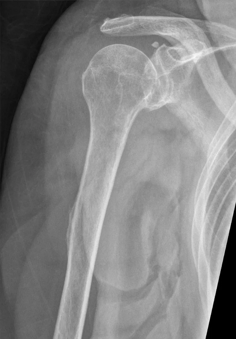
KeeperAP humerus: a keeper
Look for: The greater tubercle is in profile on the lateral side of the humeral head, so the epicondyles were parallel to the IR.
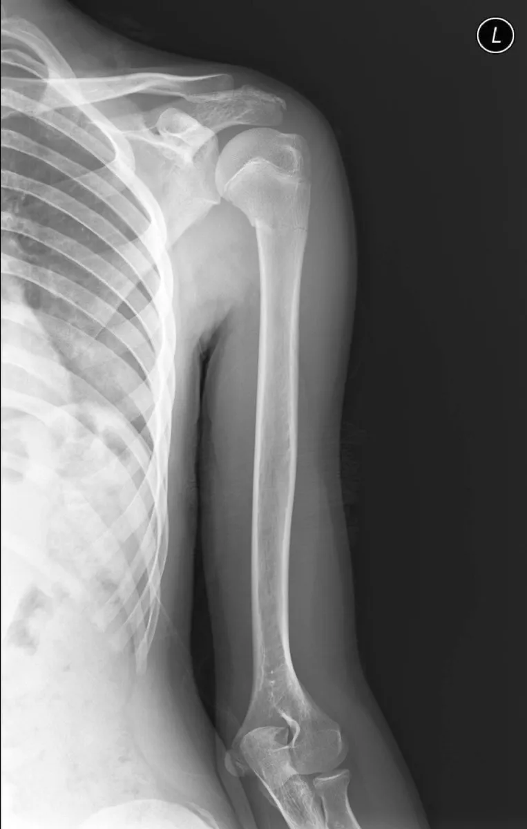
KeeperLateral humerus: a keeper
Look for: The whole humerus is on the film, from the shoulder to the elbow, with the arm turned in for the lateral.
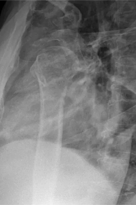
KeeperTransthoracic lateral humerus
Look for: The proximal humerus shows through the chest without the injured arm being rotated. Breathing technique blurs the ribs and lung markings over it.
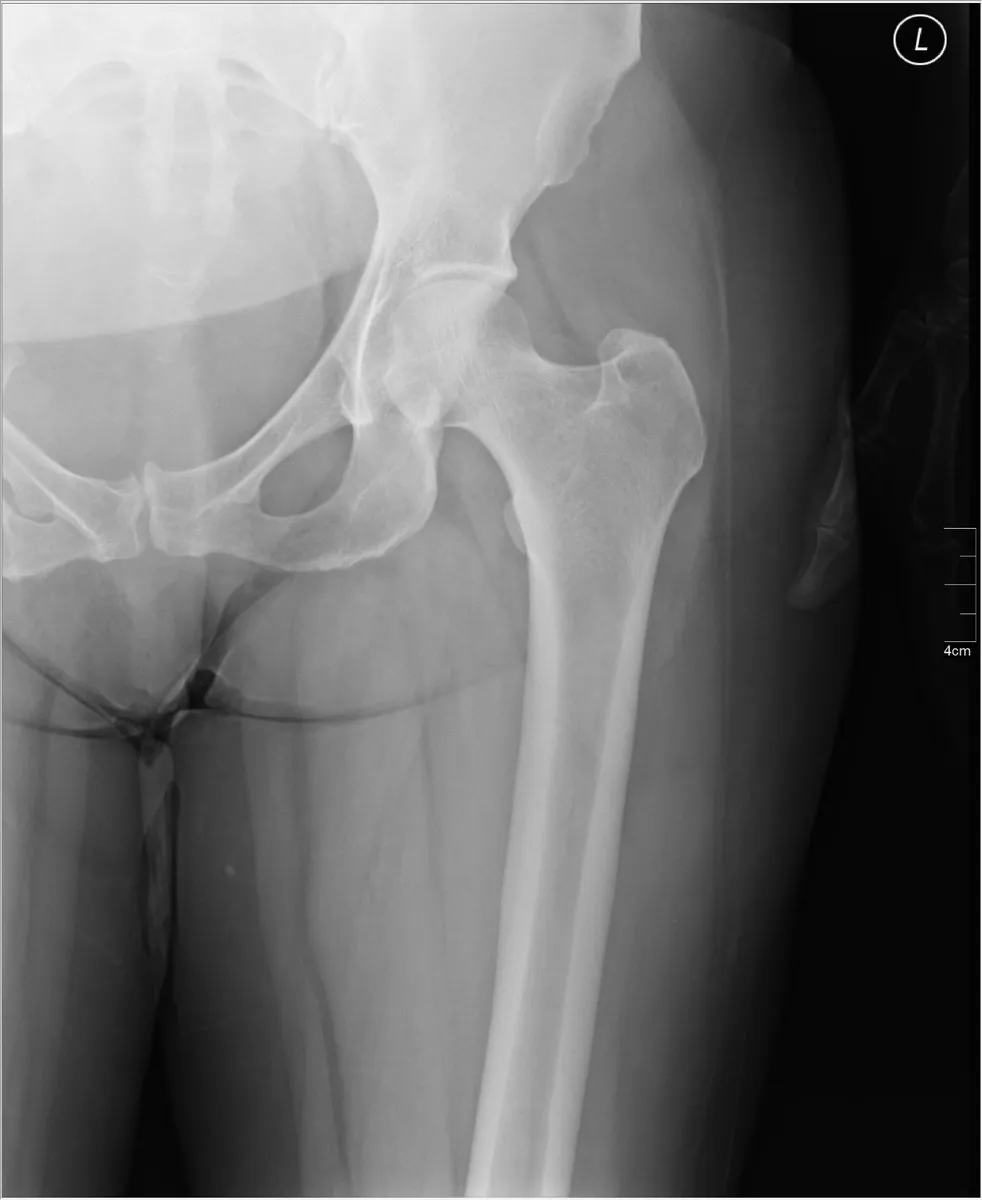
KeeperAP proximal femur: a keeper
Look for: The hip joint is included, the femoral neck shows at full length, and the lesser trochanter is small.
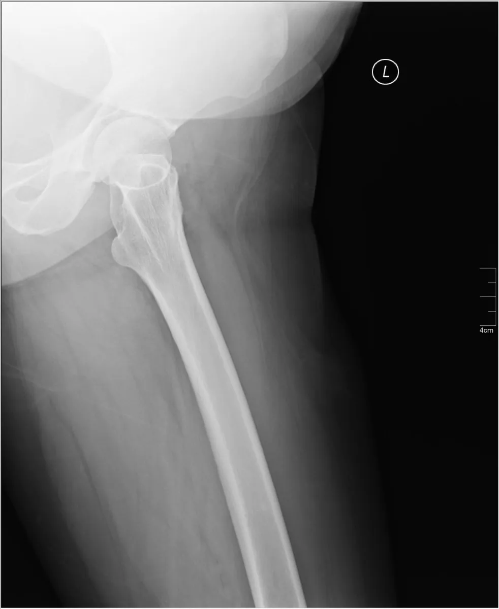
KeeperLateral proximal femur
Look for: The proximal femur is shown at a right angle to the AP, with the hip end included.
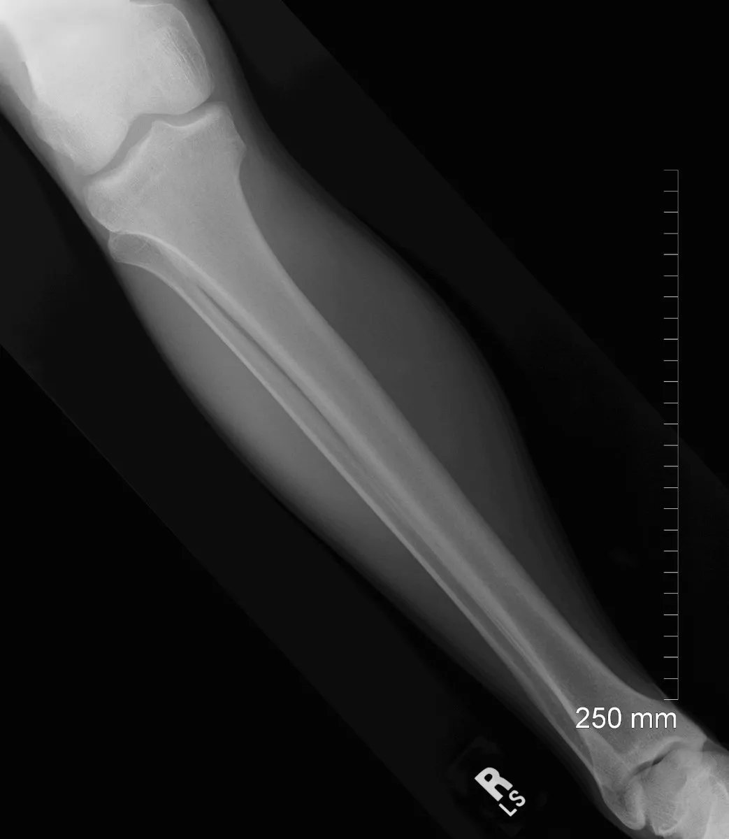
KeeperAP tibia and fibula: a keeper
Look for: The leg is placed diagonally so both the knee and the ankle fit on one IR.
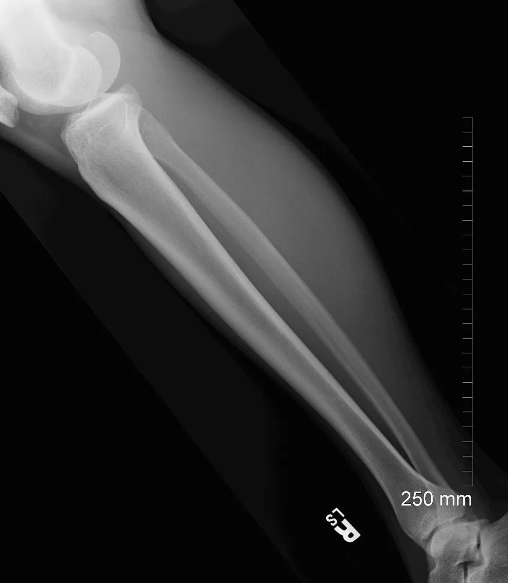
KeeperLateral tibia and fibula: a keeper
Look for: Both joints are included and the fibula lies over the back of the tibia.
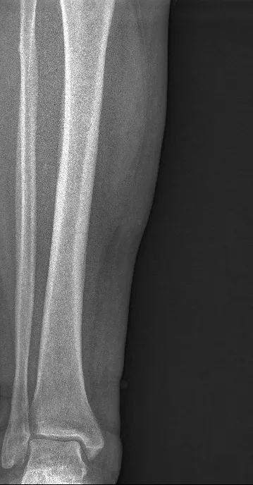
RepeatGrainy lower leg
Look for: The whole image is grainy. Too little exposure reached the detector, which is what happens when an AEC chamber is not fully under a thin part and ends the exposure early.
Fix: Center the leg over the active chamber, or switch to a manual technique for a thin extremity.
Clinical Example
A patient with a tibial shaft fracture comes in. The leg is too long for one IR the way it lies, so you place it diagonally on a 14 × 17 to include the knee and the ankle on the AP. You take the lateral cross-table without moving the leg, again with both joints.
Common Mistakes
- Leaving off the joint at the far end of the bone.
- Rotating the arm for a lateral humerus on a trauma patient.
- Taking only one image of an adult femur when it does not fit with both joints.
- Using an AEC chamber that is not fully covered by a thin limb.
Why It Matters
A fracture at one end of a long bone can hide a dislocation or a second fracture at the other end. Missing the far joint is a classic callback and a registry favorite.
Key Relationships
- Long bones: two views at right angles, both joints
- Too long for one IR → diagonal, or two overlapping images
- Cathode toward the thick end
- Trauma humerus → transthoracic lateral, no rotation
- AEC chamber not covered by the part → early termination, grainy image