
Procedures · Extremities4 min read5-question check
Knee series film check
Good knee series: open joint on the AP, condyles stacked on a lateral flexed 20 to 30°, fibular head free on the medial oblique.
The AP
On a good AP knee, the femorotibial joint space is open, the femoral condyles look symmetric, and about half of the fibular head is hidden behind the tibia. The leg is turned in about 3 to 5 degrees so the epicondyles are parallel to the IR. If the fibular head is completely hidden or completely free, or the condyles look lopsided, the leg was rotated. A narrowed joint space usually means the CR angle did not match the patient. Measure from the ASIS to the tabletop: under 19 cm angle 3 to 5 degrees caudad, 19 to 24 cm keep it perpendicular, over 24 cm angle 3 to 5 degrees cephalad.
The lateral: which edges split
On a lateral knee, the femoral condyles should stack into one outline. When they do not, look at which edges split. If the front and back edges of the condyles are offset, the knee is rotated, so fix the leg rotation until the epicondyles are perpendicular to the IR. If only the bottom edges are offset, the knee is fine and the CR angle needs adjusting. The routine is 5 to 7 degrees cephalad. The medial condyle sits farther from the IR, so it is magnified and projects lower; the angle lifts it to meet the lateral condyle. A patient with a short, wide pelvis may need more angle.
Flexion
Flex the lateral knee 20 to 30 degrees. Much more pulls the patella down into the intercondylar groove, closes the patellofemoral joint, and can hide a joint effusion. In trauma, never force flexion: a patellar fracture can be pulled apart. A knee left almost straight is the opposite miss. The condyles can stack perfectly, but it is not a standard lateral. Flexing 20 to 30 degrees relaxes the muscles around the knee and shows the most volume of the joint cavity.
Obliques and patella
The AP medial oblique, leg turned in 45 degrees, frees the fibular head and opens the proximal tibiofibular joint. The lateral oblique, leg turned out 45 degrees, puts the fibula behind the tibia. If you cannot remember which is which, look at the fibular head on the film. The tangential patella view, like the Merchant or sunrise, shows the patella in profile above the femur with the patellofemoral joint open. Rule out a patellar fracture first on the lateral.
Read the films
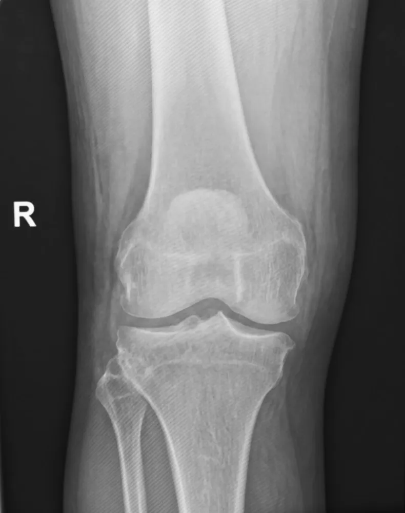
KeeperAP knee: a keeper
Look for: The femorotibial joint is open, the femoral condyles are symmetric, and about half of the fibular head is behind the tibia.
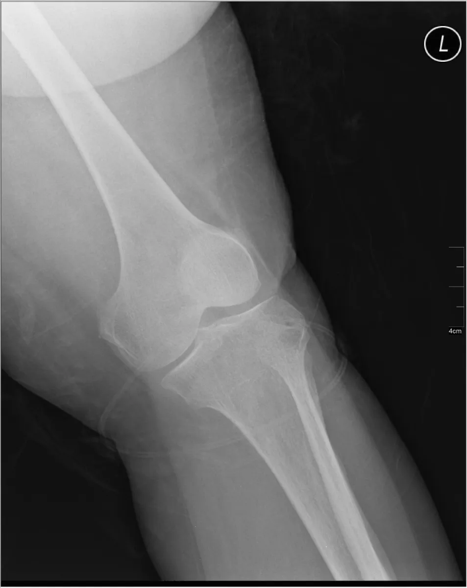
RepeatAP knee, rotated
Look for: This was ordered as an AP, but the condyles and tibia look lopsided. The leg is turned well away from a true AP.
Fix: Turn the leg in about 3 to 5 degrees so the epicondyles are parallel to the IR, with the patella centered.
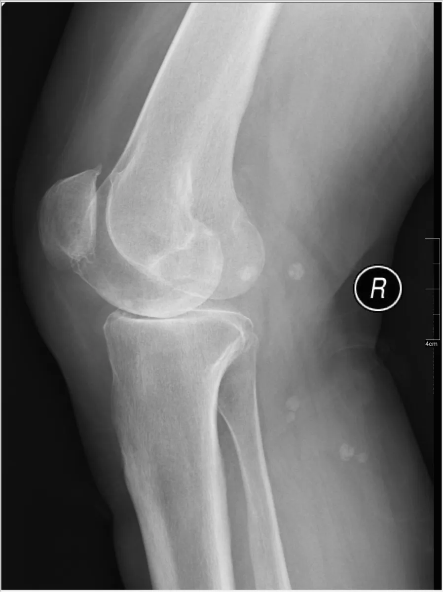
RepeatLateral knee, rotated
Look for: The condyles show as two outlines with the front and back edges offset. That means rotation, not CR angle.
Fix: Adjust the leg rotation until the epicondyles are perpendicular to the IR, then repeat.
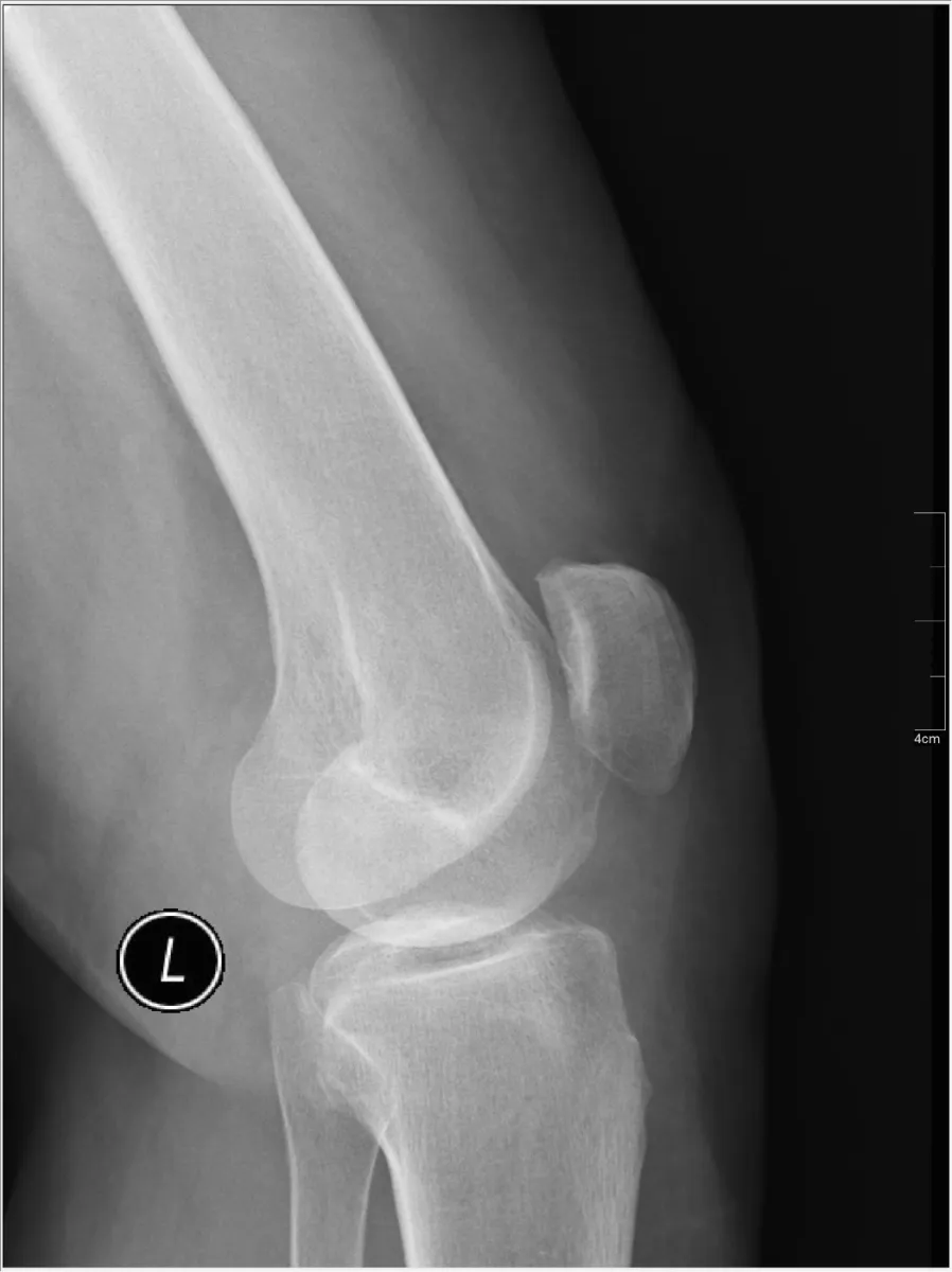
RepeatLateral knee, bottom edges split
Look for: The bottom edges of the condyles do not line up, one projecting below the other. That is a CR angle problem.
Fix: Angle 5 to 7 degrees cephalad, a little more on a patient with a wide pelvis.
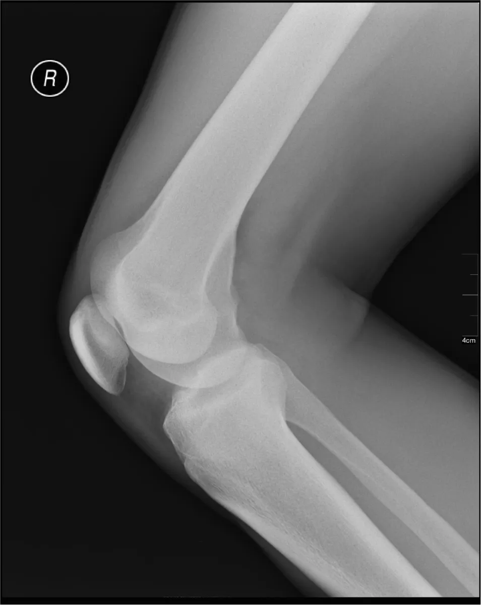
RepeatLateral knee, overflexed
Look for: The knee is bent far past 30 degrees and the patella is pulled down into the femoral groove.
Fix: Flex only 20 to 30 degrees. In trauma, take it as the patient lies and never force flexion.
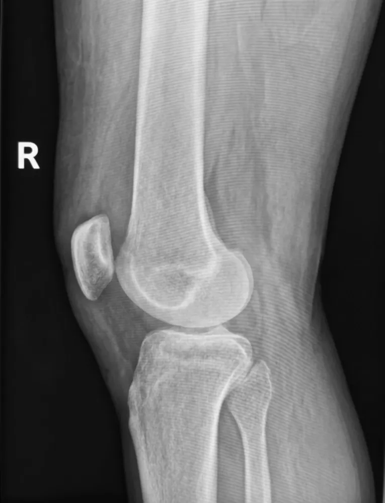
Fix next timeLateral knee, left straight
Look for: The condyles stack nicely, but the femur and tibia are almost in a straight line. The knee was not flexed.
Fix: Bend the knee 20 to 30 degrees. Good condyles alone do not make a good lateral.
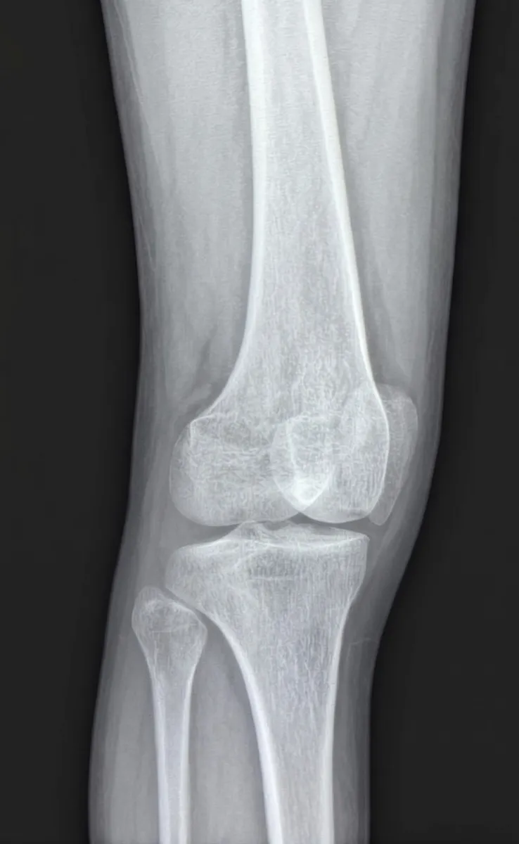
KeeperAP medial oblique: a keeper
Look for: The fibular head is free of the tibia and the proximal tibiofibular joint is open. That is how you know it is the medial oblique.
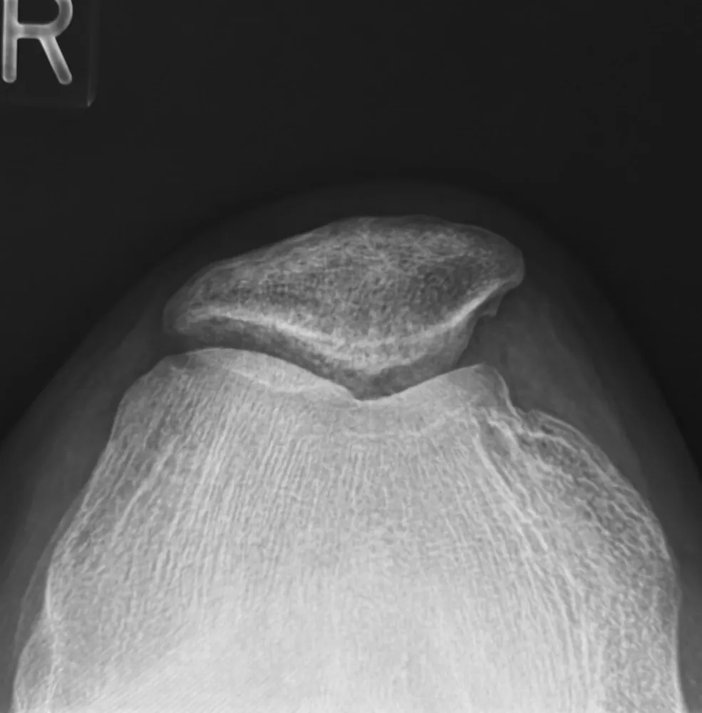
KeeperTangential patella: a keeper
Look for: The patella is in profile above the femur and the patellofemoral joint is open.
Clinical Example
Your lateral knee shows the condyles offset only along their bottom edges. Instead of rerotating the leg, you keep the position and increase the cephalad angle from 5 to 7 degrees. The repeat shows one clean condyle outline.
Common Mistakes
- Rerotating the leg when only the distal condyles are split; that is a CR angle fix.
- Flexing the lateral knee past 30 degrees, or forcing flexion in trauma.
- Using a perpendicular CR on every AP knee instead of measuring ASIS to tabletop.
- Mixing up the obliques; the medial oblique frees the fibular head.
Why It Matters
The registry loves the lateral knee: which condyle edges split and what that means. Knowing whether to fix rotation or the angle cuts repeats in half, because you change one thing instead of guessing.
Key Relationships
- Front and back condyle edges split → rotation
- Only the bottom edges split → CR angle (5 to 7° cephalad)
- Lateral flexion 20 to 30°; never forced in trauma
- AP: about half the fibular head behind the tibia
- Medial oblique: fibular head free; lateral oblique: fibula behind tibia