
Image Production · Geometry and detail2 min read5-question check
Geometric Unsharpness (Penumbra)
Edge blur from the focal spot's size. Small focal spot, short OID, and long SID keep edges sharp.
The rule
Geometric unsharpness, also called penumbra, is the blurred border around the edges of structures in the image. It happens because x-rays come from a focal spot with real width, not from a single point. Each edge is projected from many points across the focal spot, so its shadow fades instead of stopping sharply. It is present even when the patient is perfectly still.
Geometric Unsharpness
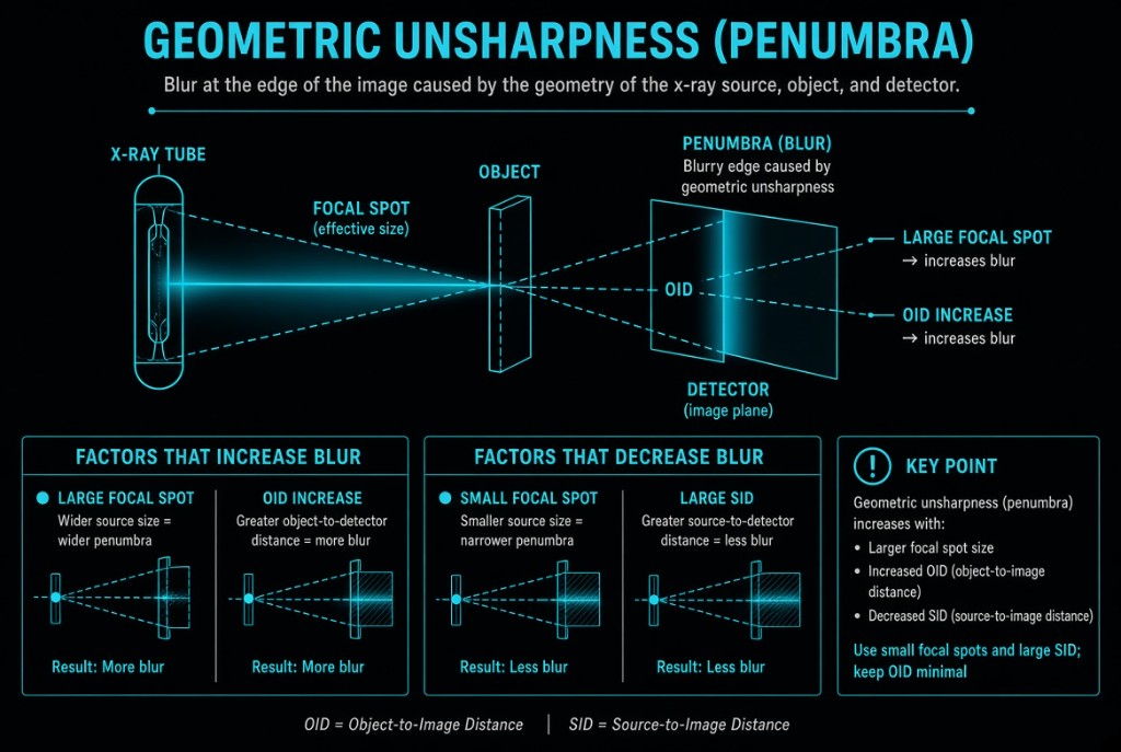
How it works
Three factors control penumbra. A larger focal spot increases it. A larger OID increases it, because the rays spread farther apart after passing the edge. A longer SID decreases it, because rays from across the focal spot arrive more nearly parallel. The anode heel effect adds a twist: the effective focal spot is smaller toward the anode, so penumbra is slightly less on the anode side of the image.
The numbers
Unsharpness equals effective focal spot size times OID divided by SOD, where SOD is SID minus OID. With a 1.0 mm focal spot, 40-inch SID, and 4-inch OID, SOD is 36 inches. Unsharpness is 1.0 × 4 ÷ 36, about 0.11 mm. Raise the SID to 72 inches with the same OID and SOD becomes 68, so unsharpness drops to about 0.06 mm. Switching to a 0.6 mm focal spot at 40 inches gives about 0.07 mm.
Reading the image
Large penumbra softens fine detail such as trabecular bone and hairline fractures, lowering spatial resolution. The small focal spot helps, but it handles less heat and limits mA, which can force a longer exposure time and invite motion blur. For most extremity work, the small focal spot and minimal OID are the right choice.
Clinical Example
A PA wrist for a suspected scaphoid fracture is done with the hand flat on the receptor, the small focal spot, and a 40-inch SID. The fine fracture line is visible. An earlier image taken with the wrist propped 3 inches above the receptor showed blurrier cortical margins.
Common Mistakes
- Confusing penumbra with motion blur, although penumbra exists even when nothing moves.
- Thinking a longer SID increases unsharpness, when it reduces it.
- Dividing by SID instead of SOD when calculating unsharpness.
- Choosing the small focal spot for a large part, then getting motion blur from the longer exposure time.
Why It Matters
Penumbra limits how fine a detail you can show, which affects whether a subtle fracture is seen. The registry tests the direction of each factor and may ask you to calculate unsharpness.
Key Relationships
- Unsharpness = focal spot size × OID ÷ SOD
- Larger focal spot → more penumbra
- Larger OID → more penumbra
- Longer SID → less penumbra
- Anode side → smaller effective focal spot → less penumbra