
Procedures · Fluoro and special studies3 min read5-question check
Foreign bodies, C-arm use and tube placement
Two views at 90 degrees locate an object. Tube under the table. An NG tip belongs below the left hemidiaphragm.
Two views
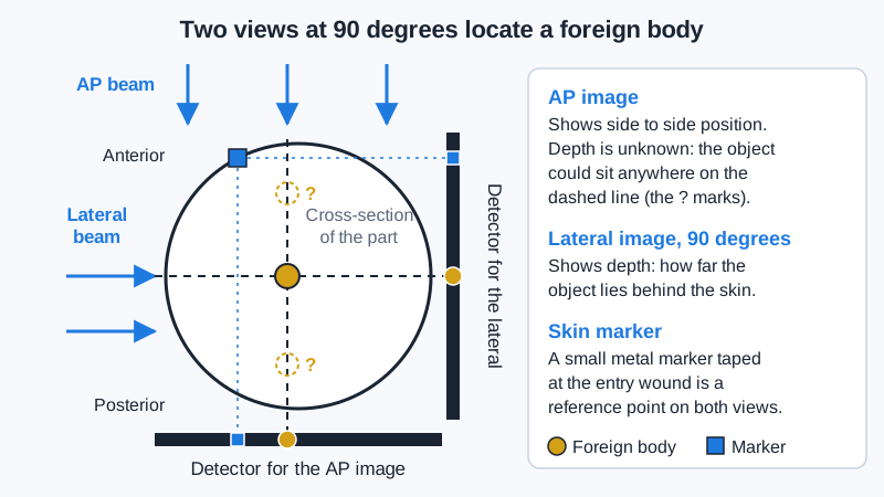
One image shows no depth, so a foreign body could lie anywhere along the beam. Localization takes at least two projections at 90 degrees, usually AP and lateral. Tape a small metal skin marker at the entry wound as a reference. For a swallowed object in a child, include the whole path from the neck through the abdomen. A coin in the esophagus usually shows as a full circle on the AP.
Two-view foreign body localization
C-arm in surgery
On a C-arm, keep the tube under the table and the receptor above, close to the patient. Scatter is strongest on the tube side, so this keeps it away from staff heads and necks. For a lateral, put the tube on the side away from the surgeon. Cover the receptor and arm with a sterile drape, and never touch the sterile field. Before starting, set the monitor orientation so the image matches the patient as the surgeon sees it. Use pulsed fluoroscopy, last-image hold and collimation, and keep at least 30 cm from source to skin. C-arms also confirm central line tips near the junction of the superior vena cava and right atrium.
Checking an enteric tube
A radiograph is the most reliable check before an enteric tube is used. Correct NG tube: it runs down the midline in the esophagus, bisects the carina, crosses the diaphragm in the midline, and ends below the left hemidiaphragm. The tip should be at least 10 cm past the gastroesophageal junction so its side holes are in the stomach. Worked example: a small-bore feeding tube turns into the right main bronchus. Feeding through it would put formula in the lung, so the nurse must hold feeds.
What to include
Include the whole tube course, from the lower neck through the upper abdomen, with both hemidiaphragms and the stomach in the field. For a tube meant to sit past the pylorus, include the duodenum. Use enough technique to see the tube through the heart, and move removable leads off its path. Report an obviously misplaced tube right away by policy.
Clinical Example
A patient on a ventilator has a new NG tube. Your portable image covers the lower neck to the upper abdomen and shows the tube bisecting the carina, crossing the diaphragm in the midline, and the tip well below the left hemidiaphragm. The radiologist clears it for use.
Common Mistakes
- Taking only one view of a foreign body and assuming its depth.
- Placing the C-arm tube above the table, which sends scatter to staff heads and necks.
- Collimating an NG tube image so tightly that the tip below the diaphragm is cut off.
- Assuming a tube is in the stomach because it reaches the level of the diaphragm.
Why It Matters
The 2027 specifications add tube placement checks, and C-arm and foreign body questions are common. Each one depends on what the radiographer includes and how the equipment is set up.
Key Relationships
- One view → no depth; two views at 90 degrees → location
- C-arm tube under the table → less scatter to staff upper body
- NG tube → bisects carina, crosses diaphragm in midline, tip below left hemidiaphragm
- NG tip → at least 10 cm past the gastroesophageal junction