
Procedures · Extremities2 min read5-question check
Foot and ankle
Angle 10 degrees toward the heel for the AP foot; rotate 15 to 20 degrees medially to open the ankle mortise.
Patient and part
For the AP foot, flex the knee and place the sole flat on the IR. For the oblique, rotate the foot 30 to 40 degrees medially. For the lateral foot, the lateral surface is down with the sole perpendicular to the IR. For the AP ankle, extend the leg and dorsiflex the foot so its long axis is vertical. For the mortise, rotate the whole leg and foot 15 to 20 degrees internally, until the intermalleolar line is parallel to the IR. The 45 degree oblique uses 45 degrees of internal rotation. The lateral ankle is taken lateral side down with the foot dorsiflexed.
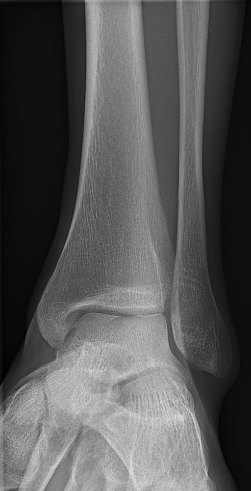
AP mortise ankle
Central ray
Use 40 inches, about 100 cm. For the AP foot, angle the CR 10 degrees posteriorly, toward the heel, so it is perpendicular to the metatarsals, and center to the base of the third metatarsal. The oblique uses the same point with a perpendicular CR. The lateral foot centers to the medial cuneiform. The AP, mortise and oblique ankle center midway between the malleoli; the lateral ankle centers to the medial malleolus. Include the base of the fifth metatarsal on the lateral ankle, because inversion injuries often fracture it.
What a good image shows
On the AP foot, the spaces between the second through fifth metatarsals are nearly equal and the tarsometatarsal joints are open. On the oblique foot, the third through fifth metatarsals are free of superimposition, and the cuboid is well seen. On the AP ankle, the medial and superior joint spaces are open, but the lateral space is normally closed by fibular overlap. On the mortise, all three joint spaces of the mortise are open and equal. The 45 degree oblique opens the distal tibiofibular joint. On a lateral foot or ankle, the fibula overlaps the posterior half of the tibia and the talar domes superimpose.
Fixing it
If the lateral mortise is still closed on a mortise view, the leg was under-rotated, so increase internal rotation toward 15 to 20 degrees. If the talar domes are not superimposed on the lateral ankle, the leg was rotated. Closed tarsometatarsal joints on the AP foot mean the CR was not perpendicular to the metatarsals.
Clinical Example
A basketball player rolls his ankle inward. You take AP, mortise and lateral ankle views, framing the lateral to include the base of the fifth metatarsal, which shows an avulsion fracture.
Common Mistakes
- Angling the AP foot CR toward the toes instead of toward the heel.
- Rotating the foot only, not the whole leg, for the mortise.
- Collimating the lateral ankle so tightly that the fifth metatarsal base is cut off.
Why It Matters
An open, even mortise lets the radiologist judge ligament injury and talar shift. The registry tests the mortise rotation, the AP foot CR angle and what each oblique shows.
Key Relationships
- AP foot: CR 10 degrees toward the heel, to base of third metatarsal
- Mortise: 15 to 20 degrees internal rotation, all spaces open
- Lateral ankle: include base of fifth metatarsal