
Image Production · Geometry and detail2 min read5-question check
Focal Spot Blooming
High mA spreads the electron stream and enlarges the effective focal spot, adding blur to fine detail.
The rule
Focal spot blooming is an increase in the actual size of the focal spot as tube current, mA, increases. The nominal focal spot size listed on the control panel, such as 0.6 mm or 1.2 mm, is measured under standard conditions. At high mA the real spot can be larger than that, and a larger effective focal spot means more blur at the edges of structures.
Focal Spot Blooming
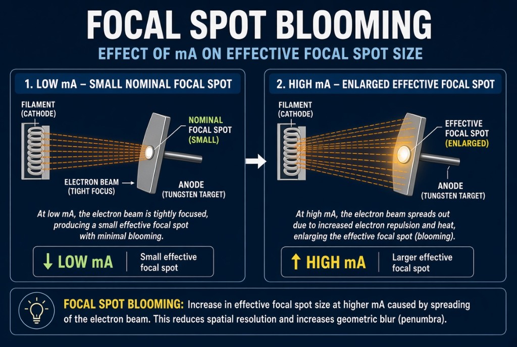
Why it happens
Electrons all carry a negative charge, so they repel each other. At high mA many more electrons stream from the filament to the anode at once, and that mutual repulsion pushes the stream wider. The negatively charged focusing cup works to keep the stream narrow, but it cannot fully overcome the crowding. Blooming tends to be greater at low kVp, because a lower voltage pulls the electrons across the tube with less force, giving them more time to spread.
The numbers
Focal spot size drives geometric unsharpness, also called penumbra. Unsharpness equals effective focal spot size times OID divided by SOD. Worked example: with a 1.2 mm focal spot, a 100 cm SID, and a 10 cm OID, SOD is 90 cm. Unsharpness is 1.2 times 10 divided by 90, or about 0.13 mm. If blooming enlarges the spot to 1.5 mm, unsharpness rises to about 0.17 mm. With zero OID, focal spot size has little effect, so blooming matters most when OID is present.
In practice
For a given mAs, you can choose a lower mA with a longer time, or a higher mA with a shorter time. Lower mA reduces blooming but increases the risk of motion blur. Choose a short time when motion is the bigger threat, as in a chest or a restless patient. Choose the small focal spot and a moderate mA when fine detail is the priority and the part can be held still, as in an extremity. The small focal spot also has lower mA limits because its heat is concentrated on a smaller target area.
Clinical Example
A PA finger is done on the small focal spot at a moderate mA and slightly longer time. A chest on a short-of-breath patient is done on the large focal spot at high mA and a very short time. Each choice controls the bigger source of blur for that exam.
Common Mistakes
- Assuming the nominal focal spot size never changes during exposure.
- Using the highest mA station by habit for small, still extremities.
- Forgetting that OID multiplies the effect of focal spot size.
- Confusing blooming with the line-focus principle, which concerns anode angle.
Why It Matters
Blooming quietly reduces spatial resolution, especially on magnified or high-OID images. The registry tests what causes it and how focal spot size, OID, and SOD combine to set unsharpness.
Key Relationships
- Increase mA → focal spot blooms larger
- Lower kVp at high mA → more blooming
- Unsharpness = focal spot size × OID ÷ SOD
- Larger effective focal spot → lower spatial resolution