
Image Production · Equipment, AEC and QC2 min read5-question check
Fluoroscopy: Image Intensifier and Flat Panel
Brightness gain is minification gain times flux gain. Magnification mode sharpens the image but raises patient dose.
The intensifier
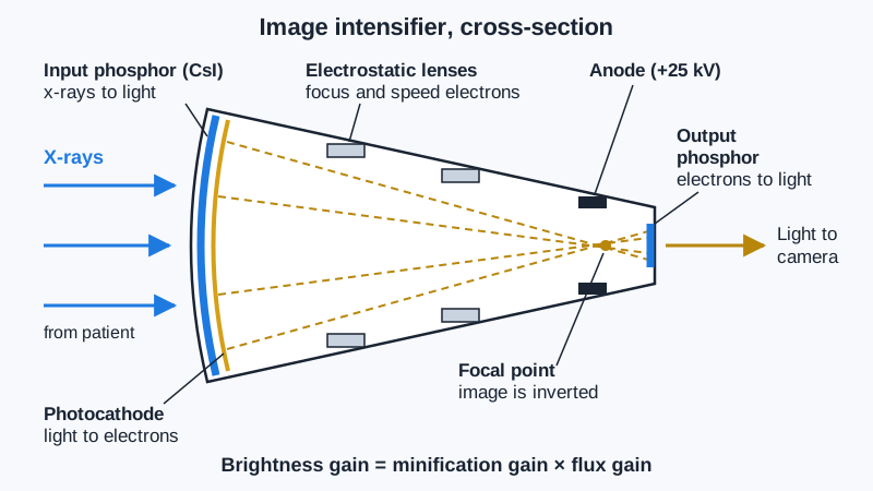
An image intensifier turns a faint x-ray pattern into a bright image. X-rays strike the input phosphor, cesium iodide, which gives off light. The photocathode, right behind it, absorbs that light and gives off electrons. Electrostatic focusing lenses steer the electrons toward the anode, which sits near the output end at about 25 kV and speeds them up. The electrons cross at a focal point and strike the small output phosphor, zinc cadmium sulfide, which gives off light for the camera.
Image intensifier, cross-section
Brightness gain
Two effects make the output image brighter. Flux gain comes from accelerating the electrons. Each electron that hits the output phosphor makes many light photons, typically about 50 times as many as the input phosphor made. Minification gain comes from squeezing the image onto a smaller phosphor. It equals the input diameter divided by the output diameter, squared. Brightness gain equals minification gain times flux gain, often several thousand.
The numbers
Worked example: a 25 cm input phosphor and a 2.5 cm output phosphor give a minification gain of (25 ÷ 2.5)², or 100. With a flux gain of 50, brightness gain is 5,000. Switching to a 17 cm magnification mode uses less of the input phosphor. The image is magnified about 1.5 times and spatial resolution improves, but minification gain drops. Automatic brightness control raises exposure to make up for it, so patient dose rises about (25 ÷ 17)², or about 2.2 times.
Flat panels and dose
Most new units use a flat-panel detector instead of an image intensifier. Flat panels have no pincushion distortion or vignetting, give uniform brightness and a rectangular field, and are smaller. Pulsed fluoroscopy fires short bursts, such as 7.5 or 15 per second, instead of a continuous beam, which cuts dose. Last-image hold keeps the final frame on the monitor with no added exposure. Also keep beam-on time short, collimate, use the least magnification needed and keep the receptor close to the patient.
Clinical Example
During a pediatric upper GI, the radiologist asks for magnification to see the duodenum. The technologist reminds the team that magnification raises dose, uses it only briefly, and sets the unit to a low pulse rate. Last-image hold lets the radiologist study each view without extra beam-on time.
Common Mistakes
- Forgetting to square the diameter ratio when finding minification gain.
- Thinking magnification mode lowers dose because the field is smaller.
- Mixing up the photocathode (light to electrons) and the output phosphor (electrons to light).
- Believing last-image hold makes a new exposure.
Why It Matters
The registry tests the order of intensifier parts, gain math and the dose effect of magnification. These same choices control patient and staff dose in every fluoro case.
Key Relationships
- Input phosphor → light → photocathode → electrons → output phosphor → light
- (Input diameter ÷ output diameter)² → minification gain
- Minification gain × flux gain → brightness gain
- Magnification mode → better resolution → higher patient dose