
Image Production · Scatter, grids and filtration2 min read5-question check
Filtration
Filtration strips useless low-energy photons, hardening the beam and cutting skin dose.
The rule
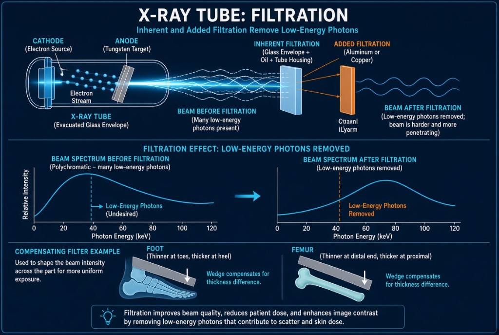
An x-ray beam leaves the target as a mix of photon energies. The lowest-energy photons have almost no chance of passing through the patient to reach the receptor. They are absorbed in the first few centimeters of tissue, adding to skin dose and contributing nothing to the image. Filtration removes them before they reach the patient. Its main purpose is reducing patient dose, especially entrance skin dose.
X-ray Filtration
Why it happens
A filter, usually aluminum, absorbs low-energy photons far more readily than high-energy ones, mainly by photoelectric absorption. The photons that remain have a higher average energy. This is called beam hardening. Filtration lowers beam quantity, the number of photons, and raises beam quality, the beam's penetrating ability. Total filtration is the sum of inherent filtration, built into the tube, and added filtration, placed in the beam.
The numbers
Radiographic units operating above 70 kVp must have at least 2.5 mm aluminum equivalent total filtration. Because filtration is hard to inspect directly, physicists verify it by measuring the half-value layer, or HVL: the thickness of absorber that cuts beam intensity in half. Worked example: a beam of 8 mGy falls to 4 mGy after one HVL, 2 mGy after two, and 1 mGy after three. A harder beam has a larger HVL. Too little filtration shows up as an HVL below the required minimum.
In practice
Filtration is part of the equipment and is checked during quality control; the radiographer does not remove it. Its effect on the image is small. The slightly harder beam lowers subject contrast a little and may need slightly more mAs for the same receptor exposure. That small cost buys a large drop in skin dose.
Clinical Example
During annual testing, the physicist measures a low HVL on a room's tube. That suggests filtration below the required minimum, so patients are getting extra skin dose from soft photons. The room is held until service corrects it.
Common Mistakes
- Saying filtration's main purpose is improving contrast.
- Thinking filtration raises beam quantity, when it lowers quantity and raises quality.
- Confusing HVL with total filtration; HVL is how filtration is checked.
- Forgetting that total filtration is inherent plus added.
Why It Matters
Filtration is one of the most effective, invisible dose reductions in radiography. The registry tests its purpose, its effect on beam quality and quantity, the 2.5 mm Al minimum, and HVL.
Key Relationships
- More filtration → lower quantity, higher quality
- Above 70 kVp → at least 2.5 mm Al total
- One HVL → intensity halved
- Total filtration = inherent + added